

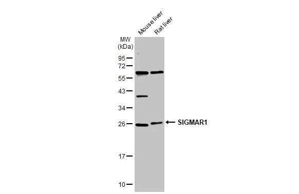
Various tissue extracts (50 μg) were separated by 12% SDS-PAGE, and the membrane was blotted with SIGMAR1 antibody [N1C3] (GTX115389) diluted at 1:1000. The HRP-conjugated anti-rabbit IgG antibody (GTX213110-01) was used to detect the primary antibody.
![Wild-type (WT) andSIGMAR1 knockout (KO) HeLa cell extracts (30 μg) were separated by 12% SDS-PAGE, and the membrane was blotted with SIGMAR1 antibody [N1C3] (GTX115389) diluted at 1:1000. The HRP-conjugated anti-rabbit IgG antibody (GTX213110-01) was used to detect the primary antibody.](https://www.genetex.com/upload/website/prouct_img/normal/GTX115389/GTX115389_44343_20210625_WB_KO_watermark_w_23060519_875.webp "Wild-type (WT) andSIGMAR1 knockout (KO) HeLa cell extracts (30 μg) were separated by 12% SDS-PAGE, and the membrane was blotted with SIGMAR1 antibody [N1C3] (GTX115389) diluted at 1:1000. The HRP-conjugated anti-rabbit IgG antibody (GTX213110-01) was used to detect the primary antibody.")
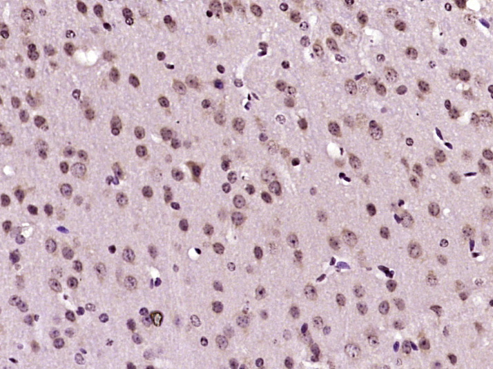
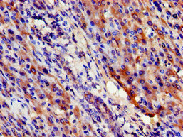
 antibody at 1:100 dilution.
Antigen Retrieval: Citrate buffer, pH 6.0, 15 min")
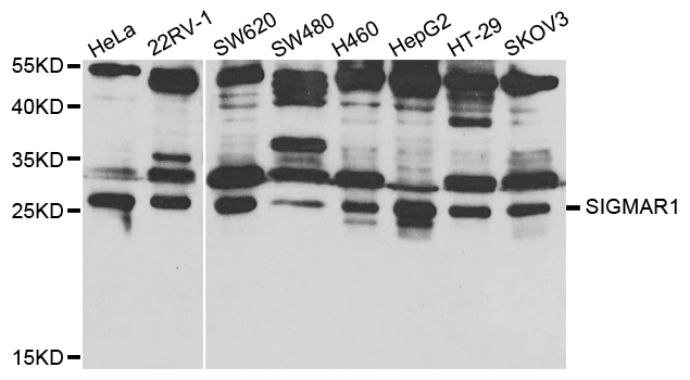
![Various whole cell extracts (30 μg) were separated by 12% SDS-PAGE, and the membrane was blotted with SIGMAR1 antibody [N1C3] (GTX115389) diluted at 1:1000. The HRP-conjugated anti-rabbit IgG antibody (GTX213110-01) was used to detect the primary antibody.](https://www.genetex.com/upload/website/prouct_img/normal/GTX115389/GTX115389_45007_20230414_WB_24120522_890.webp "Various whole cell extracts (30 μg) were separated by 12% SDS-PAGE, and the membrane was blotted with SIGMAR1 antibody [N1C3] (GTX115389) diluted at 1:1000. The HRP-conjugated anti-rabbit IgG antibody (GTX213110-01) was used to detect the primary antibody.")
Various tissue extracts (50 μg) were separated by 12% SDS-PAGE, and the membrane was blotted with SIGMAR1 antibody [N1C3] (GTX115389) diluted at 1:1000. The HRP-conjugated anti-rabbit IgG antibody (GTX213110-01) was used to detect the primary antibody.
SIGMAR1 antibody [N1C3]
GTX115389
ApplicationsWestern Blot, ImmunoHistoChemistry, ImmunoHistoChemistry Paraffin
Product group Antibodies
ReactivityHuman, Mouse, Rat, Zebra Fish
TargetSIGMAR1
Overview
- SupplierGeneTex
- Product NameSIGMAR1 antibody [N1C3]
- Delivery Days Customer9
- Application Supplier NoteWB: 1:500-1:3000. IHC-P: 1:100-1:1000. *Optimal dilutions/concentrations should be determined by the researcher.Not tested in other applications.
- ApplicationsWestern Blot, ImmunoHistoChemistry, ImmunoHistoChemistry Paraffin
- CertificationResearch Use Only
- ClonalityPolyclonal
- Concentration1.24 mg/ml
- ConjugateUnconjugated
- Gene ID10280
- Target nameSIGMAR1
- Target descriptionsigma non-opioid intracellular receptor 1
- Target synonymsALS16, DSMA2, HMNR2, OPRS1, SIG-1R, SR-BP, SR-BP1, SRBP, hSigmaR1, sigma1R, sigma non-opioid intracellular receptor 1, SR31747 binding protein 1, aging-associated gene 8 protein, sigma 1-type opioid receptor
- HostRabbit
- IsotypeIgG
- Protein IDQ99720
- Protein NameSigma non-opioid intracellular receptor 1
- Scientific DescriptionThis gene encodes a receptor protein that interacts with a variety of psychotomimetic drugs, including cocaine and amphetamines. The receptor is believed to play an important role in the cellular functions of various tissues associated with the endocrine, immune, and nervous systems. As indicated by its previous name, opioid receptor sigma 1 (OPRS1), the product of this gene was erroneously thought to function as an opioid receptor; it is now thought to be a non-opioid receptor. Alternative splicing of this gene results in transcript variants encoding distinct isoforms. [provided by RefSeq]
- ReactivityHuman, Mouse, Rat, Zebra Fish
- Storage Instruction-20°C or -80°C,2°C to 8°C
- UNSPSC41116161
Datasheet
Related products
Product group Antibodies
Anti-SIGMAR1 AntibodyA30796
ApplicationsWestern Blot, ImmunoHistoChemistry
ReactivityHuman, Mouse, Rat
- SizePrice

Product group Antibodies
Anti-Sigma1-receptor/SIGMAR1 Antibody Picoband(r)A02493-2-CARRIER-FREE
ApplicationsWestern Blot, ELISA, ImmunoHistoChemistry
ReactivityHuman, Monkey, Mouse, Rat
TargetSIGMAR1
- SizePrice
Product group Antibodies
Anti-SIGMAR1 Antibody144-05479
ApplicationsWestern Blot, ImmunoHistoChemistry
ReactivityHuman, Mouse, Rat
TargetSIGMAR1
- SizePrice

Product group Antibodies
OPRS1 Polyclonal AntibodyBS-5111R
ApplicationsImmunoFluorescence, Western Blot, ELISA, ImmunoCytoChemistry, ImmunoHistoChemistry, ImmunoHistoChemistry Frozen, ImmunoHistoChemistry Paraffin
ReactivityBovine, Canine, Human, Mouse, Porcine, Rabbit, Rat
TargetSIGMAR1
- SizePrice
Product group Antibodies
SIGMAR1 AntibodyCSB-PA021320LA01HU
ApplicationsELISA, ImmunoHistoChemistry
ReactivityHuman
TargetSIGMAR1
- SizePrice
Product group Antibodies

OPRS1 / SIGMAR1 AntibodyLS-C401334
ApplicationsWestern Blot, ELISA, ImmunoHistoChemistry
ReactivityHuman, Mouse, Rat
TargetSIGMAR1
- SizePrice

Product group Antibodies
Anti-SIGMAR1 AntibodyHPA024071
ApplicationsImmunoHistoChemistry
ReactivityHuman
TargetSIGMAR1
- SizePrice
![Various whole cell extracts (30 μg) were separated by 12% SDS-PAGE, and the membrane was blotted with SIGMAR1 antibody [HL2230] (GTX638275) diluted at 1:1000. The HRP-conjugated anti-rabbit IgG antibody (GTX213110-01) was used to detect the primary antibody.](https://www.genetex.com/upload/website/prouct_img/normal/GTX638275/GTX638275_T-44956_20230224_WB_M_R_23030219_936.webp)
Product group Antibodies
SIGMAR1 antibody [HL2230]GTX638275
ApplicationsWestern Blot, ImmunoHistoChemistry, ImmunoHistoChemistry Paraffin
ReactivityHuman, Mouse, Rat
TargetSIGMAR1
- SizePrice
![SIGMAR1 antibody [HL2274] detects SIGMAR1 protein at cell membrane by immunohistochemical analysis. Sample: Paraffin-embedded mouse stomach. SIGMAR1 stained by SIGMAR1 antibody [HL2274] (GTX638326) diluted at 1:100. Antigen Retrieval: Citrate buffer, pH 6.0, 15 min](https://www.genetex.com/upload/website/prouct_img/normal/GTX638326/GTX638326_T-44970_20230324_IHC-P_M_23032819_811.webp)
Product group Antibodies
SIGMAR1 antibody [HL2274]GTX638326
ApplicationsWestern Blot, ImmunoHistoChemistry, ImmunoHistoChemistry Paraffin
ReactivityHuman, Mouse
TargetSIGMAR1
- SizePrice