

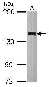
Sample (30 ug of whole cell lysate) A: HCT116 10% SDS PAGE GTX116417 diluted at 1:1000
![SIN3B antibody [N1N2-2], N-term detects SIN3B protein at nucleus by immunofluorescent analysis. Sample: HeLa cells were fixed in 4% paraformaldehyde at RT for 15 min. Green: SIN3B protein stained by SIN3B antibody [N1N2-2], N-term (GTX116417) diluted at 1:500. Red: phalloidin, a cytoskeleton marker, diluted at 1:200. Blue: Hoechst 33342 staining. Scale bar = 10 μm.](https://www.genetex.com/upload/website/prouct_img/normal/GTX116417/GTX116417_40261_20150525_IFA_w_23060519_183.webp "SIN3B antibody [N1N2-2], N-term detects SIN3B protein at nucleus by immunofluorescent analysis. Sample: HeLa cells were fixed in 4% paraformaldehyde at RT for 15 min. Green: SIN3B protein stained by SIN3B antibody [N1N2-2], N-term (GTX116417) diluted at 1:500. Red: phalloidin, a cytoskeleton marker, diluted at 1:200. Blue: Hoechst 33342 staining. Scale bar = 10 μm.")
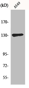
 A: mouse brain 5% SDS PAGE GTX116417 diluted at 1:500")
![Immunoprecipitation of SIN3B protein from HCT-116 whole cell extracts using 5 μg of SIN3B antibody [N1N2-2], N-term (GTX116417). Western blot analysis was performed using SIN3B antibody [N1N2-2], N-term (GTX116417) diluted at 1:500. EasyBlot anti-Rabbit IgG (GTX221666-01) was used as a secondary reagent.](https://www.genetex.com/upload/website/prouct_img/normal/GTX116417/GTX116417_40261_IP_w_23060519_942.webp "Immunoprecipitation of SIN3B protein from HCT-116 whole cell extracts using 5 μg of SIN3B antibody [N1N2-2], N-term (GTX116417). Western blot analysis was performed using SIN3B antibody [N1N2-2], N-term (GTX116417) diluted at 1:500. EasyBlot anti-Rabbit IgG (GTX221666-01) was used as a secondary reagent.")
Sample (30 ug of whole cell lysate) A: HCT116 10% SDS PAGE GTX116417 diluted at 1:1000
SIN3B antibody [N1N2-2], N-term
GTX116417
ApplicationsImmunoFluorescence, ImmunoPrecipitation, Western Blot, ImmunoCytoChemistry
Product group Antibodies
ReactivityHuman, Mouse
TargetSIN3B
Overview
- SupplierGeneTex
- Product NameSIN3B antibody [N1N2-2], N-term
- Delivery Days Customer9
- Application Supplier NoteWB: 1:500-1:3000. ICC/IF: 1:100-1:1000. IP: 1:100-1:500. *Optimal dilutions/concentrations should be determined by the researcher.Not tested in other applications.
- ApplicationsImmunoFluorescence, ImmunoPrecipitation, Western Blot, ImmunoCytoChemistry
- CertificationResearch Use Only
- ClonalityPolyclonal
- Concentration0.57 mg/ml
- ConjugateUnconjugated
- Gene ID23309
- Target nameSIN3B
- Target descriptionSIN3 transcription regulator family member B
- Target synonymspaired amphipathic helix protein Sin3b, SIN3 homolog B, transcriptional regulator, SIN3 transcription regulator homolog B, histone deacetylase complex subunit Sin3b, transcriptional corepressor Sin3b
- HostRabbit
- IsotypeIgG
- Protein IDO75182
- Protein NamePaired amphipathic helix protein Sin3b
- Scientific DescriptionActs as a transcriptional repressor. Interacts with MXI1 to repress MYC responsive genes and antagonize MYC oncogenic activities. Interacts with MAD-MAX heterodimers by binding to MAD. The heterodimer then represses transcription by tethering SIN3B to DNA. Also forms a complex with FOXK1 which represses transcription.
- ReactivityHuman, Mouse
- Storage Instruction-20°C or -80°C,2°C to 8°C
- UNSPSC41116161
Datasheet
Related products
Product group Antibodies
SIN3B AntibodyCSB-PA003323
ApplicationsImmunoFluorescence, Western Blot, ELISA
ReactivityHuman
TargetSIN3B
- SizePrice
Product group Antibodies
Anti-SIN3B AntibodyA99762
ApplicationsImmunoFluorescence, Western Blot, ELISA
ReactivityHuman
- SizePrice

Product group Antibodies
Anti-SIN3B AntibodyHPA050329
ApplicationsImmunoCytoChemistry, ImmunoHistoChemistry
ReactivityHuman
TargetSIN3B
- SizePrice
Product group Antibodies

SIN3B AntibodyLS-C497292
ApplicationsWestern Blot
ReactivityHuman, Mouse, Rat
TargetSIN3B
- SizePrice
![Immunoprecipitation of SIN3B protein from HCT-116 whole cell extracts using 5 μg of SIN3B antibody [N1N2], N-term (GTX116326). Western blot analysis was performed using SIN3B antibody [N1N2], N-term (GTX116326) diluted at 1:500. EasyBlot anti-Rabbit IgG (GTX221666-01) was used as a secondary reagent.](https://www.genetex.com/upload/website/prouct_img/normal/GTX116326/GTX116326_40282_IP_w_23060519_945.webp)
Product group Antibodies
SIN3B antibody [N1N2], N-termGTX116326
ApplicationsImmunoFluorescence, ImmunoPrecipitation, Western Blot, ImmunoCytoChemistry, ImmunoHistoChemistry, ImmunoHistoChemistry Paraffin
ReactivityHuman
TargetSIN3B
- SizePrice
Product group Antibodies
Sin3b Polyclonal AntibodyBS-4215R
ApplicationsImmunoFluorescence, Western Blot, ELISA, ImmunoCytoChemistry, ImmunoHistoChemistry, ImmunoHistoChemistry Frozen, ImmunoHistoChemistry Paraffin
ReactivityBovine, Canine, Chicken, Equine, Human, Mouse, Rat, Sheep
TargetSIN3B
- SizePrice