

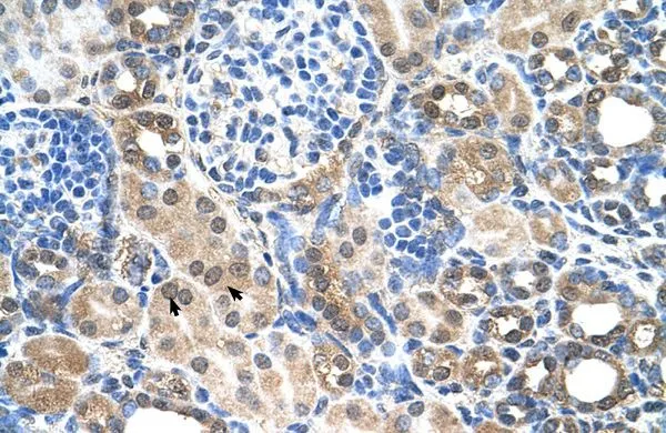
IHC-P analysis of human kidney tissue using GTX46968 SLC22A7 antibody at 4.0-8.0μg/ml.
IHC-P analysis of human kidney tissue using GTX46968 SLC22A7 antibody at 4.0-8.0μg/ml.
SLC22A7 antibody, C-term
GTX46968
ApplicationsImmunoHistoChemistry, ImmunoHistoChemistry Paraffin
Product group Antibodies
ReactivityHuman
TargetSLC22A7
Overview
- SupplierGeneTex
- Product NameSLC22A7 antibody, C-term
- Delivery Days Customer9
- Application Supplier NoteIHC-P: 2-10 ug/ml. *Optimal dilutions/concentrations should be determined by the researcher.Not tested in other applications.
- ApplicationsImmunoHistoChemistry, ImmunoHistoChemistry Paraffin
- CertificationResearch Use Only
- ClonalityPolyclonal
- Concentration0.5-1 mg/ml
- ConjugateUnconjugated
- Gene ID10864
- Target nameSLC22A7
- Target descriptionsolute carrier family 22 member 7
- Target synonymsNLT, OAT2, hOAT11, solute carrier family 22 member 7, hOAT2, liver-specific transporter, novel liver transporter, organic anion transporter 11, organic anion transporter 2, solute carrier family 22 (organic anion transporter), member 7
- HostRabbit
- IsotypeIgG
- Protein IDQ9Y694
- Protein NameSolute carrier family 22 member 7
- Scientific DescriptionThe protein encoded by this gene is involved in the sodium-independent transport and excretion of organic anions, some of which are potentially toxic. The encoded protein is an integral membrane protein and appears to be localized to the basolateral membrane of the kidney. Alternatively spliced transcript variants encoding different isoforms have been described. [provided by RefSeq, Jul 2008]
- ReactivityHuman
- Storage Instruction-20°C or -80°C,2°C to 8°C
- UNSPSC41116161
Datasheet
Related products

Product group Antibodies
Anti-OAT2/SLC22A7 Antibody Picoband(r)A05273-2-CARRIER-FREE
ApplicationsFlow Cytometry, Western Blot, ELISA, ImmunoHistoChemistry
ReactivityHuman, Mouse, Rat
TargetSLC22A7
- SizePrice
Product group Antibodies
Anti-SLC22A7 Antibody144-60932
ApplicationsWestern Blot
ReactivityHuman, Mouse, Rat
TargetSLC22A7
- SizePrice
Product group Antibodies

SLC22A7 / OAT2 AntibodyLS-C750108
ApplicationsWestern Blot
ReactivityHuman, Mouse, Rat
TargetSLC22A7
- SizePrice
Product group Antibodies
SLC22A7 Polyclonal AntibodyBS-19808R
ApplicationsImmunoFluorescence, Western Blot, ELISA, ImmunoCytoChemistry, ImmunoHistoChemistry, ImmunoHistoChemistry Frozen, ImmunoHistoChemistry Paraffin
ReactivityBovine, Equine, Human, Mouse, Porcine, Rabbit, Rat
TargetSLC22A7
- SizePrice

Product group Antibodies
Anti-SLC22A7 AntibodyHPA030220
ApplicationsImmunoHistoChemistry
ReactivityHuman
TargetSLC22A7
- SizePrice
Product group Antibodies
SLC22A7 antibody, N-termGTX47048
ApplicationsWestern Blot
ReactivityHuman
TargetSLC22A7
- SizePrice
Product group Antibodies
SLC22A7 antibody, N-termGTX47049
ApplicationsWestern Blot, ImmunoHistoChemistry, ImmunoHistoChemistry Paraffin
ReactivityHuman
TargetSLC22A7
- SizePrice
Product group Antibodies
Anti-SLC22A7 AntibodyCAB15137
ApplicationsWestern Blot, ELISA
ReactivityHuman
TargetSLC22A7
- SizePrice