

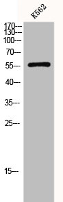
SLC2A1 / GLUT-1 Antibody (aa92-492)
LS-C407646
ApplicationsWestern Blot, ImmunoCytoChemistry, ImmunoHistoChemistry, ImmunoHistoChemistry Frozen, ImmunoHistoChemistry Paraffin
Product group Antibodies
ReactivityHuman, Mouse, Rat
TargetSLC2A1
Overview
- SupplierLifeSpan BioSciences
- Product NameSLC2A1 / GLUT-1 Antibody (aa92-492)
- Delivery Days Customer23
- Application Supplier NoteWB: The detection limit for SLC2A1 is approximately 0.1 ng/lane under reducing conditions. IHC: Antigen retrieval by boiling the paraffin sections in 10 mM citrate buffer, pH6.0, for 20 minutes is required for the staining of formalin/paraffin sections.. ICC (0.5 - 1 µg/ml), IHC, IHC-Fr (0.5 - 1 µg/ml), IHC-P (0.5 - 1 µg/ml), WB (0.1 - 0.5 µg/ml) WB: The detection limit for SLC2A1 is approximately 0.1 ng/lane under reducing conditions. IHC: Antigen retrieval by boiling the paraffin sections in 10 mM citrate buffer, pH6.0, for 20 minutes is required for the staining of formalin/paraffin sections.
- ApplicationsWestern Blot, ImmunoCytoChemistry, ImmunoHistoChemistry, ImmunoHistoChemistry Frozen, ImmunoHistoChemistry Paraffin
- CertificationResearch Use Only
- ClonalityPolyclonal
- ConjugateUnconjugated
- Estimated Purity...
- Gene ID6513
- Target nameSLC2A1
- Target descriptionsolute carrier family 2 member 1
- Target synonymsCSE, DYT17, DYT18, DYT9, EIG12, GLUT, GLUT-1, GLUT1, GLUT1DS, HTLVR, PED, SDCHCN, solute carrier family 2, facilitated glucose transporter member 1, choreoathetosis/spasticity, episodic (paroxysmal choreoathetosis/spasticity), dystonia gene 18, dystonia gene 9, glucose transporter type 1, erythrocyte/brain, hepG2 glucose transporter, human T-cell leukemia virus (I and II) receptor, receptor for HTLV-1 and HTLV-2, solute carrier family 2 (facilitated glucose transporter), member 1
- HostRabbit
- ReactivityHuman, Mouse, Rat
- Storage Instruction-20°C,2°C to 8°C
- UNSPSC41116161
Related products
Product group Antibodies
SLC2A1 AntibodyCSB-PA002728
ApplicationsWestern Blot, ELISA, ImmunoHistoChemistry
ReactivityHuman, Mouse, Rat
TargetSLC2A1
- SizePrice
Product group Antibodies
Anti-GLUT1 AntibodyA95873
ApplicationsWestern Blot, ELISA, ImmunoHistoChemistry
ReactivityHuman, Mouse, Rat
- SizePrice

Product group Antibodies
Anti-SLC2A1 AntibodyHPA031345
ApplicationsImmunoCytoChemistry, ImmunoHistoChemistry
ReactivityHuman, Mouse
TargetSLC2A1
- SizePrice
Product group Antibodies

SLC2A1 / GLUT-1 AntibodyLS-C402347
ApplicationsWestern Blot, ELISA, ImmunoHistoChemistry
ReactivityHuman, Mouse, Rat
TargetSLC2A1
- SizePrice
Product group Antibodies
Slc2A1 Polyclonal AntibodyCAC10560
ApplicationsWestern Blot, ELISA, ImmunoHistoChemistry
ReactivityMouse
TargetSLC2A1
- SizePrice
Product group Antibodies
anti-GLUT1, pAb (IN116)AG-25B-0040
ApplicationsImmunoPrecipitation, Western Blot, ImmunoHistoChemistry
ReactivityHuman, Mouse, Rat
TargetSLC2A1
- SizePrice

Product group Antibodies
Anti-Glucose Transporter GLUT1/SLC2A1 Antibody Picoband(r)PB9435-CARRIER-FREE
ApplicationsFlow Cytometry, ImmunoFluorescence, Western Blot, ImmunoCytoChemistry, ImmunoHistoChemistry, ImmunoHistoChemistry Frozen
ReactivityHuman, Mouse, Rat
TargetSLC2A1
- SizePrice
![IHC-P analysis of human cerebrum (grey matter) tissue using GTX04469 GluT1 antibody [MSVA-401R] HistoMAX?. A particularly strong GluT1 staining of endothelial cells is seen in the brain.](https://www.genetex.com/upload/website/prouct_img/normal/GTX04469/GTX04469_20230728_IHC-P_56_23072722_577.webp)
Product group Antibodies
ApplicationsImmunoHistoChemistry, ImmunoHistoChemistry Paraffin
ReactivityHuman
TargetSLC2A1
- SizePrice
Product group Antibodies
References
GLUT1 Polyclonal Antibodybs-0472R
ApplicationsFlow Cytometry, ImmunoFluorescence, Western Blot, ELISA, ImmunoCytoChemistry, ImmunoHistoChemistry, ImmunoHistoChemistry Frozen, ImmunoHistoChemistry Paraffin
ReactivityBovine, Canine, Chicken, Human, Mouse, Porcine, Rat, Sheep
TargetSLC2A1
- SizePrice