

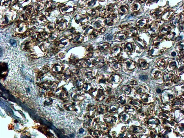
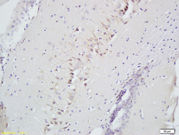
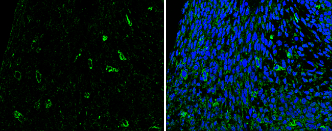
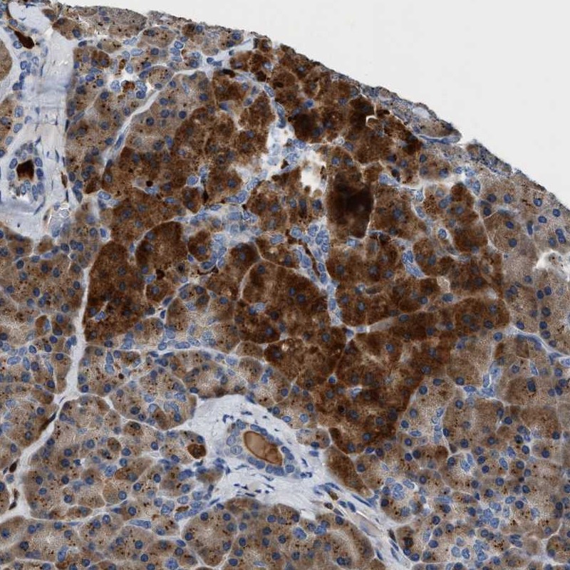
IHC-P analysis of human spinal cord using GTX89329 SLIT2 antibody, Internal. Antigen retrieval : Tris/EDTA buffer pH 9 Dilution : 4μg/ml
IHC-P analysis of human spinal cord using GTX89329 SLIT2 antibody, Internal. Antigen retrieval : Tris/EDTA buffer pH 9 Dilution : 4μg/ml
SLIT2 antibody, Internal
GTX89329
ApplicationsImmunoHistoChemistry, ImmunoHistoChemistry Paraffin
Product group Antibodies
ReactivityHuman
TargetSLIT2
Overview
- SupplierGeneTex
- Product NameSLIT2 antibody, Internal
- Delivery Days Customer9
- Application Supplier NoteIHC-P: 4-8microg/ml. *Optimal dilutions/concentrations should be determined by the researcher.Not tested in other applications.
- ApplicationsImmunoHistoChemistry, ImmunoHistoChemistry Paraffin
- CertificationResearch Use Only
- ClonalityPolyclonal
- Concentration0.50 mg/ml
- ConjugateUnconjugated
- Gene ID9353
- Target nameSLIT2
- Target descriptionslit guidance ligand 2
- Target synonymsSLIL3, Slit-2, slit homolog 2 protein
- HostGoat
- IsotypeIgG
- Protein IDO94813
- Protein NameSlit homolog 2 protein
- Scientific DescriptionThis gene encodes a member of the slit family of secreted glycoproteins, which are ligands for the Robo family of immunoglobulin receptors. Slit proteins play highly conserved roles in axon guidance and neuronal migration and may also have functions during other cell migration processes including leukocyte migration. Members of the slit family are characterized by an N-terminal signal peptide, four leucine-rich repeats, nine epidermal growth factor repeats, and a C-terminal cysteine knot. Proteolytic processing of this protein gives rise to an N-terminal fragment that contains the four leucine-rich repeats and five epidermal growth factor repeats and a C-terminal fragment that contains four epidermal growth factor repeats and the cysteine knot. Both full length and cleaved proteins are secreted extracellularly and can function in axon repulsion as well as other specific processes. Alternative splicing results in multiple transcript variants. [provided by RefSeq, Sep 2015]
- ReactivityHuman
- Storage Instruction-20°C or -80°C,2°C to 8°C
- UNSPSC41116161
Datasheet
Related products
Product group Antibodies
Anti-SLIT2 AntibodyA46292
ApplicationsImmunoHistoChemistry
ReactivityHuman, Mouse
- SizePrice

Product group Antibodies
Anti-SLIT2 Antibody Picoband(r)A01627-1-CARRIER-FREE
ApplicationsFlow Cytometry, Western Blot, ELISA
ReactivityHuman
TargetSLIT2
- SizePrice
Product group Antibodies
SLIT2 Polyclonal AntibodyBS-2743R
ApplicationsImmunoFluorescence, Western Blot, ELISA, ImmunoCytoChemistry, ImmunoHistoChemistry, ImmunoHistoChemistry Frozen, ImmunoHistoChemistry Paraffin
ReactivityHuman, Mouse, Rat
TargetSLIT2
- SizePrice
Product group Antibodies
SLIT2 AntibodyCSB-PA136864
ApplicationsWestern Blot, ELISA, ImmunoHistoChemistry
ReactivityHuman, Mouse, Rat
TargetSLIT2
- SizePrice
Product group Antibodies

Goat anti-SLIT2EB07287
ApplicationsELISA, ImmunoHistoChemistry
ReactivityCanine, Human, Mouse, Rat
TargetSLIT2
- SizePrice
Product group Antibodies
ApplicationsImmunoPrecipitation, Western Blot, ImmunoCytoChemistry, ImmunoHistoChemistry
ReactivityMouse, Rat
TargetSLIT2
- SizePrice
Product group Antibodies

SLIT2 AntibodyLS-C403581
ApplicationsWestern Blot, ELISA, ImmunoHistoChemistry
ReactivityHuman, Mouse, Rat
TargetSLIT2
- SizePrice
Product group Antibodies
SLIT2 antibodyGTX118220
ApplicationsWestern Blot, ImmunoHistoChemistry, ImmunoHistoChemistry Paraffin
ReactivityHuman, Mouse, Rat
TargetSLIT2
- SizePrice
Product group Antibodies
Anti-SLIT2 AntibodyHPA019511
ApplicationsImmunoHistoChemistry
ReactivityHuman
TargetSLIT2
- SizePrice