

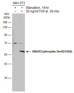
Untreated (–) and treated (+) NIH-3T3 whole cell extracts (30 μg) were separated by 10% SDS-PAGE, and the membrane was blotted with SMAD3 (phospho Ser423/425) antibody (GTX129841) diluted at 1:500. The HRP-conjugated anti-rabbit IgG antibody (GTX213110-01) was used to detect the primary antibody.
 and treated (+) HeLa whole cell extracts (30 μg) were separated by 12% SDS-PAGE, and the membrane was blotted with SMAD3 (phospho Ser423/425) antibody (GTX129841) diluted at 1:500.")
Untreated (–) and treated (+) NIH-3T3 whole cell extracts (30 μg) were separated by 10% SDS-PAGE, and the membrane was blotted with SMAD3 (phospho Ser423/425) antibody (GTX129841) diluted at 1:500. The HRP-conjugated anti-rabbit IgG antibody (GTX213110-01) was used to detect the primary antibody.
SMAD3 (phospho Ser423/425) antibody
GTX129841
ApplicationsWestern Blot
Product group Antibodies
ReactivityHuman, Mouse
TargetSMAD3
Overview
- SupplierGeneTex
- Product NameSMAD3 (phospho Ser423/425) antibody
- Delivery Days Customer9
- Application Supplier NoteWB: 1:500-1:3000. *Optimal dilutions/concentrations should be determined by the researcher.Not tested in other applications.
- ApplicationsWestern Blot
- CertificationResearch Use Only
- ClonalityPolyclonal
- Concentration1 mg/ml
- ConjugateUnconjugated
- Gene ID4088
- Target nameSMAD3
- Target descriptionSMAD family member 3
- Target synonymsHSPC193, HsT17436, JV15-2, LDS1C, LDS3, MADH3, hMAD-3, hSMAD3, mad3, mothers against decapentaplegic homolog 3, MAD homolog 3, MAD, mothers against decapentaplegic homolog 3, SMA- and MAD-related protein 3, SMAD, mothers against DPP homolog 3, mad homolog JV15-2, mad protein homolog, mothers against DPP homolog 3
- HostRabbit
- IsotypeIgG
- Protein IDP84022
- Protein NameMothers against decapentaplegic homolog 3
- Scientific DescriptionThe protein encoded by this gene belongs to the SMAD, a family of proteins similar to the gene products of the Drosophila gene mothers against decapentaplegic (Mad) and the C. elegans gene Sma. SMAD proteins are signal transducers and transcriptional modulators that mediate multiple signaling pathways. This protein functions as a transcriptional modulator activated by transforming growth factor-beta and is thought to play a role in the regulation of carcinogenesis. [provided by RefSeq]
- ReactivityHuman, Mouse
- Storage Instruction-20°C or -80°C,2°C to 8°C
- UNSPSC12352203
References
- Huang C, Lu HF, Chen YH, et al. Curcumin, demethoxycurcumin, and bisdemethoxycurcumin induced caspase-dependent and -independent apoptosis via Smad or Akt signaling pathways in HOS cells. BMC Complement Med Ther. 2020,20(1):68. doi: 10.1186/s12906-020-2857-1Read this paper
- Hsieh WY, Chang TH, Chang HF, et al. Renal chymase-dependent pathway for angiotensin II formation mediated acute kidney injury in a mouse model of aristolochic acid I-induced acute nephropathy. PLoS One. 2019,14(1):e0210656. doi: 10.1371/journal.pone.0210656Read this paper
Datasheet
Related products
Product group Antibodies

SMAD3-S208 AntibodyABX034610
ApplicationsFlow Cytometry, ImmunoFluorescence, Western Blot, ELISA, ImmunoCytoChemistry, ImmunoHistoChemistry
- SizePrice
Product group Antibodies
Anti-SMAD3 Antibody144-07536
ApplicationsImmunoFluorescence, Western Blot, ImmunoHistoChemistry
ReactivityHuman, Mouse, Rat
TargetSMAD3
- SizePrice
![Untreated (–) and treated (+) A549 whole cell extracts (30 μg) were separated by 10% SDS-PAGE, and the membrane was blotted with SMAD3 (phospho Ser423/Ser425) antibody [GT1207] (GTX00969) diluted at 1:500. The HRP-conjugated anti-rabbit IgG antibody (GTX213110-01) was used to detect the primary antibody.](https://www.genetex.com/upload/website/prouct_img/normal/GTX00969/GTX00969_4000000170_20200313_WB_treatment_TGF-beta1_w_23053121_737.webp)
Product group Antibodies
References
ApplicationsWestern Blot, ImmunoHistoChemistry, ImmunoHistoChemistry Paraffin
ReactivityHuman, Mouse, Rat
TargetSMAD3
- SizePrice
Product group Antibodies
References
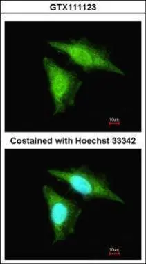
SMAD2 / SMAD3 antibodyGTX111123
ApplicationsFlow Cytometry, ImmunoFluorescence, ImmunoPrecipitation, Western Blot, ChIP Chromatin ImmunoPrecipitation, ImmunoCytoChemistry, ImmunoHistoChemistry, ImmunoHistoChemistry Paraffin
ReactivityHuman, Mouse, Rat
TargetSMAD3
- SizePrice
Product group Antibodies
SMAD3 (phospho Ser425) antibodyGTX78942
ApplicationsImmunoHistoChemistry, ImmunoHistoChemistry Paraffin
ReactivityHuman
TargetSMAD3
- SizePrice
Product group Antibodies
SMAD3 Polyclonal AntibodyCAC13933
ApplicationsWestern Blot, ELISA
TargetSMAD3
- SizePrice
Product group Antibodies
References
ApplicationsFlow Cytometry, ImmunoFluorescence, Western Blot, ELISA, ImmunoCytoChemistry, ImmunoHistoChemistry, ImmunoHistoChemistry Frozen, ImmunoHistoChemistry Paraffin
ReactivityBovine, Canine, Chicken, Equine, Human, Mouse, Porcine, Rat
TargetSMAD3
- SizePrice
Product group Antibodies
Phospho-SMAD3(S425) Monoclonal AntibodyCSB-MA388929
ApplicationsELISA, ImmunoHistoChemistry
ReactivityHuman, Mouse, Rat
TargetSMAD3
- SizePrice