

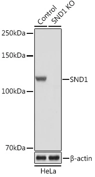
WB analysis of normal (control) and knockout (KO) HeLa cell lysate using GTX33508 SND1 antibody. Dilution : 1:3000 Loading : 25μg per lane


WB analysis of normal (control) and knockout (KO) HeLa cell lysate using GTX33508 SND1 antibody. Dilution : 1:3000 Loading : 25μg per lane
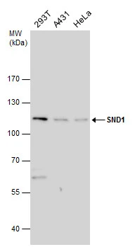
SND1 antibody
GTX33508
ApplicationsImmunoFluorescence, ImmunoPrecipitation, Western Blot, ImmunoCytoChemistry, ImmunoHistoChemistry, ImmunoHistoChemistry Paraffin
Product group Antibodies
ReactivityHuman, Mouse, Rat
TargetSND1
Overview
- SupplierGeneTex
- Product NameSND1 antibody
- Delivery Days Customer9
- Application Supplier NoteWB: 1:500 - 1:2000. ICC/IF: 1:50 - 1:200. IHC-P: 1:50 - 1:200. IP: 1:50 - 1:200. *Optimal dilutions/concentrations should be determined by the researcher.Not tested in other applications.
- ApplicationsImmunoFluorescence, ImmunoPrecipitation, Western Blot, ImmunoCytoChemistry, ImmunoHistoChemistry, ImmunoHistoChemistry Paraffin
- CertificationResearch Use Only
- ClonalityPolyclonal
- ConjugateUnconjugated
- Gene ID27044
- Target nameSND1
- Target descriptionstaphylococcal nuclease and tudor domain containing 1
- Target synonymsTDRD11, TSN, Tudor-SN, p100, staphylococcal nuclease domain-containing protein 1, EBNA2 coactivator p100, testis tissue sperm-binding protein Li 82P, tudor domain-containing protein 11
- HostRabbit
- IsotypeIgG
- Protein IDQ7KZF4
- Protein NameStaphylococcal nuclease domain-containing protein 1
- Scientific DescriptionThis gene encodes a transcriptional co-activator that interacts with the acidic domain of Epstein-Barr virus nuclear antigen 2 (EBNA 2), a transcriptional activator that is required for B-lymphocyte transformation. Other transcription factors that interact with this protein are signal transducers and activators of transcription, STATs. This protein is also thought to be essential for normal cell growth. A similar protein in mammals and other organisms is a component of the RNA-induced silencing complex (RISC). [provided by RefSeq, Jul 2016]
- ReactivityHuman, Mouse, Rat
- Storage Instruction-20°C or -80°C,2°C to 8°C
- UNSPSC41116161
Datasheet
Related products
Product group Antibodies
Anti-SND1 AntibodyA31235
ApplicationsImmunoFluorescence, Western Blot, ImmunoHistoChemistry
ReactivityHuman, Mouse, Rat
- SizePrice

Product group Antibodies
Anti-SND1 Antibody Picoband(r)A02602-3-CARRIER-FREE
ApplicationsFlow Cytometry, ImmunoFluorescence, Western Blot, ELISA, ImmunoCytoChemistry, ImmunoHistoChemistry
ReactivityHuman, Mouse, Rat
TargetSND1
- SizePrice
Product group Antibodies
Anti-SND1 Antibody144-05874
ApplicationsImmunoFluorescence, Western Blot, ImmunoHistoChemistry
ReactivityHuman, Mouse, Rat
TargetSND1
- SizePrice
Product group Antibodies
SND1 AntibodyCSB-PA004724
ApplicationsWestern Blot, ELISA
ReactivityHuman, Mouse, Rat
TargetSND1
- SizePrice
![WB analysis of HeLa (1), Jukat (2), HepG2 (3) SMMC-7721 (4) cell lysate using GTX83194 SND1 antibody [2D7].](https://www.genetex.com/upload/website/prouct_img/normal/GTX83194/GTX83194_20170912_WB_w_23061322_322.webp)
Product group Antibodies
SND1 antibody [2D7]GTX83194
ApplicationsWestern Blot, ELISA
ReactivityHuman
TargetSND1
- SizePrice

Product group Antibodies
Anti-SND1 AntibodyHPA002529
ApplicationsWestern Blot, ImmunoHistoChemistry
ReactivityHuman, Mouse, Rat
TargetSND1
- SizePrice
Product group Antibodies
SND1 antibodyGTX130135
ApplicationsImmunoPrecipitation, Western Blot
ReactivityHuman
TargetSND1
- SizePrice
Product group Antibodies
SND1 antibodyGTX130136
ApplicationsWestern Blot
ReactivityHuman
TargetSND1
- SizePrice
Product group Antibodies

SND1 AntibodyLS-C748465
ApplicationsWestern Blot, ImmunoHistoChemistry
ReactivityHuman, Mouse, Rat
TargetSND1
- SizePrice