

SNRPD2 Antibody
LS-C748409
ApplicationsImmunoFluorescence, Western Blot
Product group Antibodies
ReactivityHuman, Mouse, Rat
TargetSNRPD2
Overview
- SupplierLifeSpan BioSciences
- Product NameSNRPD2 Antibody
- Delivery Days Customer23
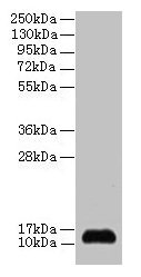
- Application Supplier NoteThe predicted MW is 12kDa/13kDa, while the observed MW by Western blot was 14kDa.
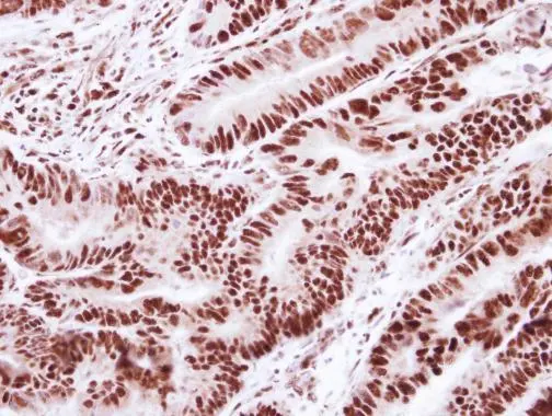
- ApplicationsImmunoFluorescence, Western Blot
- Applications SupplierIF (1:50 - 1:200), WB (1:500 - 1:2000) The predicted MW is 12kDa/13kDa, while the observed MW by Western blot was 14kDa.
- CertificationResearch Use Only
- ClonalityPolyclonal
- ConjugateUnconjugated
- Estimated Purity...
- Gene ID6633
- Target nameSNRPD2
- Target descriptionsmall nuclear ribonucleoprotein D2 polypeptide
- Target synonymsSMD2, SNRPD1, Sm-D2, small nuclear ribonucleoprotein Sm D2, small nuclear ribonucleoprotein D2 polypeptide 16.5kDa, snRNP core protein D2
- HostRabbit
- IsotypeIgG
- ReactivityHuman, Mouse, Rat
- Storage Instruction-20°C
- UNSPSC12352203
Related products

Product group Antibodies
Anti-SNRPD2 Antibody Picoband(r)A12026-1-CARRIER-FREE
ApplicationsFlow Cytometry, ImmunoFluorescence, Western Blot, ELISA, ImmunoCytoChemistry, ImmunoHistoChemistry
ReactivityHuman, Mouse, Rat
TargetSNRPD2
- SizePrice
Product group Antibodies
Anti-SNRPD2 AntibodyA31793
ApplicationsImmunoFluorescence, Western Blot, ImmunoHistoChemistry
ReactivityHuman, Mouse, Rat
- SizePrice
Product group Antibodies
SNRPD2 AntibodyCSB-PA02635A0RB
ApplicationsWestern Blot, ELISA
ReactivityHuman
TargetSNRPD2
- SizePrice

Product group Antibodies
Anti-SNRPD2 AntibodyHPA041437
ApplicationsWestern Blot, ImmunoCytoChemistry, ImmunoHistoChemistry
ReactivityHuman, Mouse, Rat
TargetSNRPD2
- SizePrice
Product group Antibodies
SNRPD2 Polyclonal AntibodyCAC14003
ApplicationsWestern Blot, ELISA
TargetSNRPD2
- SizePrice
Product group Antibodies
SNRPD2 antibodyGTX101846
ApplicationsImmunoFluorescence, Western Blot, ImmunoCytoChemistry, ImmunoHistoChemistry, ImmunoHistoChemistry Paraffin
ReactivityHuman, Mouse
TargetSNRPD2
- SizePrice
Product group Antibodies
Anti-SNRPD2 Antibody144-06983
ApplicationsImmunoFluorescence, Western Blot, ImmunoHistoChemistry
ReactivityHuman, Mouse, Rat
TargetSNRPD2
- SizePrice