

IHC-P analysis of human spleen tissue using GTX31617 SNRPN antibody. Working concentration : 20 μg/ml

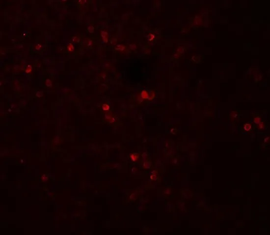
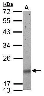
 0.5 and (B) 1 μg/ml")
IHC-P analysis of human spleen tissue using GTX31617 SNRPN antibody. Working concentration : 20 μg/ml
SNRPN antibody
GTX31617
ApplicationsWestern Blot, ELISA, ImmunoHistoChemistry, ImmunoHistoChemistry Paraffin
Product group Antibodies
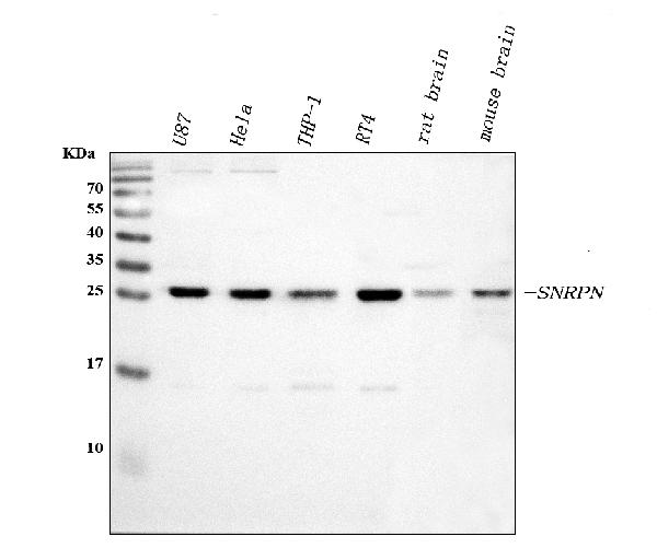
ReactivityHuman, Mouse, Rat
TargetSNRPN
Overview
- SupplierGeneTex
- Product NameSNRPN antibody
- Delivery Days Customer9
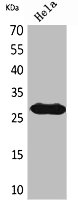
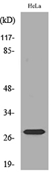
- Application Supplier NoteWB: 0.5 - 1 microg/mL. IHC-P: 5 microg/mL. *Optimal dilutions/concentrations should be determined by the researcher.Not tested in other applications.
- ApplicationsWestern Blot, ELISA, ImmunoHistoChemistry, ImmunoHistoChemistry Paraffin
- CertificationResearch Use Only
- ClonalityPolyclonal
- Concentration1 mg/ml
- ConjugateUnconjugated
- Gene ID6638
- Target nameSNRPN
- Target descriptionsmall nuclear ribonucleoprotein polypeptide N
- Target synonymsHCERN3, PWCR, RT-LI, SM-D, SMN, SNRNP-N, SNURF-SNRPN, sm-N, small nuclear ribonucleoprotein-associated protein N, SM protein N, sm protein D, tissue-specific splicing protein
- HostRabbit
- IsotypeIgG
- Protein IDP63162
- Protein NameSmall nuclear ribonucleoprotein-associated protein N
- Scientific DescriptionThe protein encoded by this gene is one polypeptide of a small nuclear ribonucleoprotein complex and belongs to the snRNP SMB/SMN family. The protein plays a role in pre-mRNA processing, possibly tissue-specific alternative splicing events. Although individual snRNPs are believed to recognize specific nucleic acid sequences through RNA-RNA base pairing, the specific role of this family member is unknown. The protein arises from a bicistronic transcript that also encodes a protein identified as the SNRPN upstream reading frame (SNURF). Multiple transcription initiation sites have been identified and extensive alternative splicing occurs in the 5 untranslated region. Additional splice variants have been described but sequences for the complete transcripts have not been determined. The 5 UTR of this gene has been identified as an imprinting center. Alternative splicing or deletion caused by a translocation event in this paternally-expressed region is responsible for Angelman syndrome or
- ReactivityHuman, Mouse, Rat
- Storage Instruction-20°C or -80°C,2°C to 8°C
- UNSPSC41116161
Datasheet
Related products
Product group Antibodies
SNRPN AntibodyCSB-PA011056
ApplicationsWestern Blot, ELISA
ReactivityHuman, Mouse, Rat
TargetSNRPN
- SizePrice
Product group Antibodies
Anti-SNRPN AntibodyA97213
ApplicationsWestern Blot, ELISA
ReactivityHuman, Mouse, Rat
- SizePrice
Product group Antibodies

SNRPN Antibody (21-70 aa, Internal)LS-C387208
ApplicationsWestern Blot, ELISA
ReactivityHuman, Mouse, Rat
TargetSNRPN
- SizePrice
Product group Antibodies
Anti-SNRPN Antibody Picoband(r)PB9441-CARRIER-FREE
ApplicationsImmunoFluorescence, Western Blot, ImmunoCytoChemistry, ImmunoHistoChemistry
ReactivityHamster, Human, Mouse, Rat
TargetSNRPN
- SizePrice
Product group Antibodies
SNURF antibody, InternalGTX82489
ApplicationsImmunoHistoChemistry, ImmunoHistoChemistry Paraffin
ReactivityHuman
TargetSNRPN
- SizePrice
Product group Antibodies
SNRPN antibodyGTX114927
ApplicationsWestern Blot, ImmunoHistoChemistry, ImmunoHistoChemistry Paraffin
ReactivityHuman, Mouse, Rat
TargetSNRPN
- SizePrice
Product group Antibodies
Anti-SNRPN Antibody144-61169
ApplicationsWestern Blot
ReactivityHuman, Mouse, Rat
TargetSNRPN
- SizePrice
Product group Antibodies
SNRPN Monoclonal AntibodyBSM-60533M
ApplicationsWestern Blot
ReactivityHuman, Mouse, Rat
TargetSNRPN
- SizePrice