

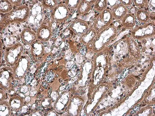
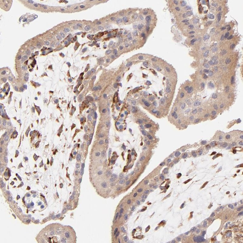
SPARC antibody detects SPARC protein in rat kidney by immunohistochemical analysis. Sample: Paraffin-embedded rat kidney. SPARC antibody (GTX133747) diluted at 1:500.
Antigen Retrieval: Citrate buffer, pH 6.0, 15 min
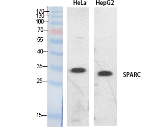
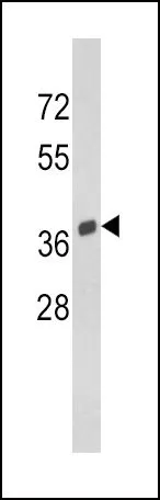
 was separated by 12% SDS-PAGE, and the membrane was blotted with SPARC antibody (GTX133747) diluted at 1:1000. The HRP-conjugated anti-rabbit IgG antibody (GTX213110-01) was used to detect the primary antibody.")
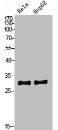
 was separated by 12% SDS-PAGE, and the membranes were blotted with SPARC antibody (GTX133747) diluted at 1:1000 and competitor's antibody (sc-25574) diluted at 1:1000. The HRP-conjugated anti-rabbit IgG antibody (GTX213110-01) was used to detect the primary antibody.")
 diluted at 1:500, and competitor's antibody diluted at 1:500.
Antigen Retrieval: Citrate buffer, pH 6.0, 15 min")
![SPARC antibody detects SPARC protein by immunohistochemical analysis. Samples: Paraffin-embedded mouse retina. Green: SPARC protein stained by SPARC antibody (GTX133747) diluted at 1:250. Red: beta Tubulin 3/ Tuj1, a marker, stained by beta Tubulin 3/ Tuj1 antibody [GT1338] (GTX631831) diluted at 1:500. Blue: Fluoroshield with DAPI (GTX30920).
Antigen Retrieval: Citrate buffer, pH 6.0, 15 min](https://www.genetex.com/upload/website/prouct_img/normal/GTX133747/GTX133747_42970_20171127_IHC-P_M_w_23060523_317.webp "SPARC antibody detects SPARC protein by immunohistochemical analysis. Samples: Paraffin-embedded mouse retina. Green: SPARC protein stained by SPARC antibody (GTX133747) diluted at 1:250. Red: beta Tubulin 3/ Tuj1, a marker, stained by beta Tubulin 3/ Tuj1 antibody [GT1338] (GTX631831) diluted at 1:500. Blue: Fluoroshield with DAPI (GTX30920).
Antigen Retrieval: Citrate buffer, pH 6.0, 15 min")
 diluted at 1:500.
Antigen Retrieval: Citrate buffer, pH 6.0, 15 min")

SPARC antibody detects SPARC protein in rat kidney by immunohistochemical analysis. Sample: Paraffin-embedded rat kidney. SPARC antibody (GTX133747) diluted at 1:500.
Antigen Retrieval: Citrate buffer, pH 6.0, 15 min
SPARC antibody
GTX133747
ApplicationsWestern Blot, ImmunoHistoChemistry, ImmunoHistoChemistry Paraffin
Product group Antibodies
ReactivityHuman, Mouse, Rat
TargetSPARC
Overview
- SupplierGeneTex
- Product NameSPARC antibody
- Delivery Days Customer9
- Application Supplier NoteWB: 1:500-1:3000. IHC-P: 1:100-1:1000. *Optimal dilutions/concentrations should be determined by the researcher.Not tested in other applications.
- ApplicationsWestern Blot, ImmunoHistoChemistry, ImmunoHistoChemistry Paraffin
- CertificationResearch Use Only
- ClonalityPolyclonal
- Concentration0.54 mg/ml
- ConjugateUnconjugated
- Gene ID6678
- Target nameSPARC
- Target descriptionsecreted protein acidic and cysteine rich
- Target synonymsBM-40, OI17, ON, ONT, SPARC, basement-membrane protein 40, secreted protein, acidic, cysteine-rich (osteonectin)
- HostRabbit
- IsotypeIgG
- Protein IDP09486
- Protein NameSPARC
- Scientific DescriptionSecreted protein acidic and rich in cysteine/osteonectin/BM40, or SPARC, is a matrix-associated protein that elicits changes in cell shape, inhibits cell-cycle progression, and influences the synthesis of extracellular matrix (ECM) (Bradshaw et al., 2003 [PubMed 12721366]).[supplied by OMIM]
- ReactivityHuman, Mouse, Rat
- Storage Instruction-20°C or -80°C,2°C to 8°C
- UNSPSC41116161
Datasheet
Related products
Product group Antibodies
Anti-SPARC AntibodyA97211
ApplicationsWestern Blot, ELISA
ReactivityHuman, Mouse, Rat
- SizePrice

Product group Antibodies
Anti-SPARC Antibody Picoband(r)A00862-1-CARRIER-FREE
ApplicationsWestern Blot
ReactivityHuman, Mouse, Rat
TargetSPARC
- SizePrice
Product group Antibodies
Anti-SPARC Antibody144-60680
ApplicationsWestern Blot
ReactivityHuman
TargetSPARC
- SizePrice
Product group Antibodies
SPARC Polyclonal AntibodyBS-1133R
ApplicationsImmunoFluorescence, Western Blot, ELISA, ImmunoCytoChemistry, ImmunoHistoChemistry, ImmunoHistoChemistry Frozen, ImmunoHistoChemistry Paraffin
ReactivityBovine, Canine, Equine, Human, Mouse, Porcine, Rabbit, Rat
TargetSPARC
- SizePrice
Product group Antibodies
SPARC AntibodyCSB-PA006034
ApplicationsWestern Blot, ELISA
ReactivityHuman, Mouse, Rat
TargetSPARC
- SizePrice
Product group Antibodies
ApplicationsImmunoPrecipitation, Western Blot, ImmunoCytoChemistry, ImmunoHistoChemistry
ReactivityMouse, Porcine, Rat
TargetSPARC
- SizePrice
Product group Antibodies

Osteonectin / SPARC Antibody (aa18-303)LS-C401048
ApplicationsWestern Blot, ELISA, ImmunoHistoChemistry
ReactivityHuman, Mouse, Rat
TargetSPARC
- SizePrice
Product group Antibodies
SPARC antibody, C-termGTX81749
ApplicationsFlow Cytometry, Western Blot, ImmunoHistoChemistry, ImmunoHistoChemistry Paraffin
ReactivityHuman
TargetSPARC
- SizePrice
Product group Antibodies
Anti-SPARC AntibodyHPA002989
ApplicationsWestern Blot, ImmunoHistoChemistry
ReactivityHuman
TargetSPARC
- SizePrice