

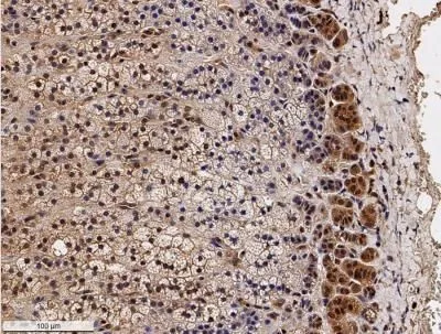
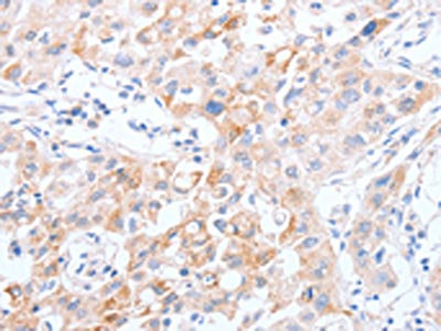
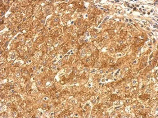
IHC-P analysis of human adrenal gland tissue using GTX30467 SR-BI antibody. Dilution : 1:100


IHC-P analysis of human adrenal gland tissue using GTX30467 SR-BI antibody. Dilution : 1:100
SR-BI antibody
GTX30467
ApplicationsFlow Cytometry, ImmunoFluorescence, ImmunoPrecipitation, Western Blot, ImmunoCytoChemistry, ImmunoHistoChemistry, ImmunoHistoChemistry Frozen, ImmunoHistoChemistry Paraffin, Neutralisation/Blocking
Product group Antibodies
ReactivityHuman, Mouse, Rat
Overview
- SupplierGeneTex
- Product NameSR-BI antibody
- Delivery Days Customer9
- Application Supplier NoteWB: 1:500. ICC/IF: 1:50 - 1:200. IHC-P: 1:100. IP: 1:100. Neutralizing/Inhibition: 1:500 - 1000. *Optimal dilutions/concentrations should be determined by the researcher.Not tested in other applications.
- ApplicationsFlow Cytometry, ImmunoFluorescence, ImmunoPrecipitation, Western Blot, ImmunoCytoChemistry, ImmunoHistoChemistry, ImmunoHistoChemistry Frozen, ImmunoHistoChemistry Paraffin, Neutralisation/Blocking
- CertificationResearch Use Only
- ClonalityPolyclonal
- ConjugateUnconjugated
- HostRabbit
- IsotypeIgG
- Scientific DescriptionHigh density lipoproteins (HDLs) play a critical role in cholesterol metabolism and their plasma concentrations are inversely correlated with risk for atherosclerosis. The SR-BI binds HDLs and mediates selective uptake of HDL cholesteryl ester. SR-BI binds HDL with high affinity, is expressed primarily in liver and nonplacental steroidgenic tissues, and mediates selective cholesterol uptake by a distinct mechanism. In mice, it seems that SR-BI plays a key role in determining the levels of plasma lipoprotein cholesterol and the accumulation of cholesterol stores in the adrenal gland.
- ReactivityHuman, Mouse, Rat
- Storage Instruction-20°C or -80°C,2°C to 8°C
- UNSPSC41116161
References
- Characterization of hepatitis C virus particle subpopulations reveals multiple usage of the scavenger receptor BI for entry steps. Dao Thi VL et al., 2012 Sep 7, J Biol ChemRead this paper
Datasheet
Related products
Product group Antibodies
SCARB1 AntibodyCSB-PA166219
ApplicationsELISA, ImmunoHistoChemistry
ReactivityHuman
TargetSCARB1
- SizePrice

Product group Antibodies
Anti-Scavenging Receptor SR-BI/SCARB1 Antibody Picoband(r)A01093-1-CARRIER-FREE
ApplicationsFlow Cytometry, Western Blot, ELISA
ReactivityHuman, Mouse
TargetSCARB1
- SizePrice
Product group Antibodies

Anti-CD36 [185-1G2 (B467)]Ab01539-1.1
ApplicationsFlow Cytometry, ImmunoFluorescence, ImmunoPrecipitation, ImmunoHistoChemistry, Neutralisation/Blocking
ReactivityHuman
TargetSCARB1
- SizePrice
Product group Antibodies
Anti-SCARB1 AntibodyA29805
ApplicationsWestern Blot, ImmunoHistoChemistry
ReactivityHuman, Mouse, Rat
- SizePrice

Product group Antibodies
Anti-SCARB1 AntibodyHPA066285
ApplicationsImmunoCytoChemistry
ReactivityHuman
TargetSCARB1
- SizePrice
Product group Antibodies

Goat anti-SCARB1 / SR-BIEB12300
ApplicationsWestern Blot, ELISA, ImmunoHistoChemistry
ReactivityHuman
TargetSCARB1
- SizePrice
Product group Antibodies

SCARB1 / SR-BI AntibodyLS-C400786
ApplicationsELISA, ImmunoHistoChemistry
ReactivityHuman
TargetSCARB1
- SizePrice
Product group Antibodies
Scarb1 Polyclonal AntibodyCAC10509
ApplicationsELISA, ImmunoHistoChemistry
TargetSCARB1
- SizePrice
Product group Antibodies
SR-BI antibodyGTX113645
ApplicationsImmunoFluorescence, Western Blot, ImmunoCytoChemistry, ImmunoHistoChemistry, ImmunoHistoChemistry Paraffin
ReactivityHuman, Mouse, Rat
TargetSCARB1
- SizePrice