

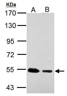
Sample (30 μg of whole cell lysate)? ? A: JC ? B: C2C12 ? 7.5% SDS PAGE? ? GTX113555 diluted at 1:10000 ? The HRP-conjugated anti-rabbit IgG antibody (GTX213110-01) was used to detect the primary antibody.
 antibody at 1:100 dilution.
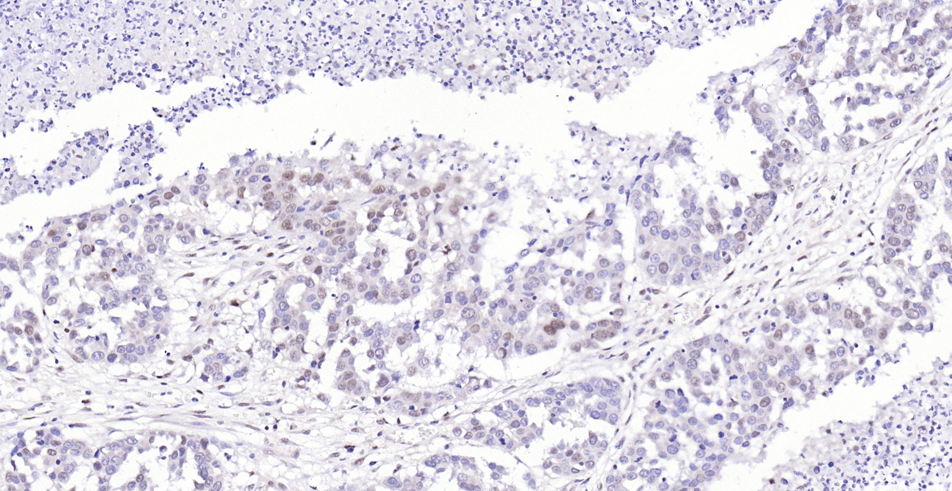
Antigen Retrieval: Trilogy? (EDTA based, pH 8.0) buffer, 15min")
 dilution: 1:1000.
Antigen Retrieval: Trilogy? (EDTA based, pH 8.0) buffer, 15min")
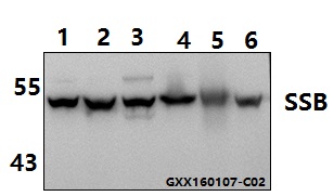
 antibody at 1:200 dilution.")
 dilution: 1:1000.
Antigen Retrieval: Trilogy? (EDTA based, pH 8.0) buffer, 15min")
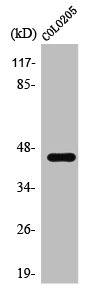
 A: 293T 10% SDS PAGE GTX113555 diluted at 1:1000 The HRP-conjugated anti-rabbit IgG antibody (GTX213110-01) was used to detect the primary antibody.")
Sample (30 μg of whole cell lysate)? ? A: JC ? B: C2C12 ? 7.5% SDS PAGE? ? GTX113555 diluted at 1:10000 ? The HRP-conjugated anti-rabbit IgG antibody (GTX213110-01) was used to detect the primary antibody.
SSB antibody
GTX113555
ApplicationsImmunoFluorescence, Western Blot, ImmunoCytoChemistry, ImmunoHistoChemistry, ImmunoHistoChemistry Paraffin
Product group Antibodies
ReactivityHuman, Mouse, Rat
TargetSSB
Overview
- SupplierGeneTex
- Product NameSSB antibody
- Delivery Days Customer9
- Application Supplier NoteWB: 1:1000-1:10000. ICC/IF: 1:100-1:1000. IHC-P: 1:100-1:1000. *Optimal dilutions/concentrations should be determined by the researcher.Not tested in other applications.
- ApplicationsImmunoFluorescence, Western Blot, ImmunoCytoChemistry, ImmunoHistoChemistry, ImmunoHistoChemistry Paraffin
- CertificationResearch Use Only
- ClonalityPolyclonal
- Concentration0.52 mg/ml
- ConjugateUnconjugated
- Gene ID6741
- Target nameSSB
- Target descriptionsmall RNA binding exonuclease protection factor La
- Target synonymsLARP3, La, La/SSB, lupus La protein, La ribonucleoprotein domain family, member 3, SS-B, SS-B/La protein, Sjogren syndrome antigen B, autoantigen La, la autoantigen, lupus La antigen, sjoegren syndrome type B antigen
- HostRabbit
- IsotypeIgG
- Protein IDP05455
- Protein NameLupus La protein
- Scientific DescriptionLa is involved in diverse aspects of RNA metabolism, including binding and protecting 3-prime UUU(OH) elements of newly RNA polymerase III (see MIM 606007)-transcribed RNA, processing 5-prime and 3-prime ends of pre-tRNA precursors, acting as an RNA chaperone, and binding viral RNAs associated with hepatitis C virus. La protein was originally defined by its reactivity with autoantibodies from patients with Sjogren syndrome (MIM 270150) and systemic lupus erythematosus (SLE; MIM 152700) (Teplova et al., 2006 [PubMed 16387655]).[supplied by OMIM]
- ReactivityHuman, Mouse, Rat
- Storage Instruction-20°C or -80°C,2°C to 8°C
- UNSPSC41116161
Datasheet
Related products
Product group Antibodies
Anti-SSB (T362) AntibodyA24948
ApplicationsWestern Blot
ReactivityHuman, Mouse, Rat
- SizePrice

Product group Antibodies
Anti-SSB Antibody Picoband(r)A00705-1-CARRIER-FREE
ApplicationsFlow Cytometry, ImmunoFluorescence, Western Blot, ELISA, ImmunoCytoChemistry, ImmunoHistoChemistry
ReactivityHuman
TargetSSB
- SizePrice
Product group Antibodies
Anti-SSB Antibody144-00630
ApplicationsImmunoFluorescence, Western Blot, ImmunoHistoChemistry
ReactivityHuman
TargetSSB
- SizePrice
Product group Antibodies
SSB Polyclonal AntibodyBS-0276R
ApplicationsImmunoFluorescence, ELISA, ImmunoCytoChemistry, ImmunoHistoChemistry, ImmunoHistoChemistry Frozen, ImmunoHistoChemistry Paraffin
ReactivityHuman
TargetSSB
- SizePrice
Product group Antibodies
SSB AntibodyCSB-PA004161
ApplicationsWestern Blot, ELISA, ImmunoHistoChemistry
ReactivityHuman
TargetSSB
- SizePrice
Product group Antibodies
SSB Polyclonal AntibodyCAC13943
ApplicationsWestern Blot, ELISA
ReactivityMouse
TargetSSB
- SizePrice
Product group Antibodies

SSB / La AntibodyLS-C403611
ApplicationsWestern Blot, ELISA, ImmunoHistoChemistry
ReactivityHuman
TargetSSB
- SizePrice
![IHC-P analysis of human prostate tissue using GTX83557 SSB antibody [9D6]. Antigen retrieval : Heat-induced epitope retrieval by 10mM citrate buffer, pH6.0, 100oC for 10min.](https://www.genetex.com/upload/website/prouct_img/normal/GTX83557/GTX83557_1612_IHC-P_w_23061420_655.webp)
Product group Antibodies
SSB antibody [9D6]GTX83557
ApplicationsFlow Cytometry, ImmunoFluorescence, ImmunoPrecipitation, Western Blot, ImmunoCytoChemistry, ImmunoHistoChemistry, ImmunoHistoChemistry Paraffin
ReactivityCanine, Human, Monkey
TargetSSB
- SizePrice
![WB analysis of various samples using GTX83558 SSB antibody [1E11]. Loading : 10 ug per lane Dilution : 1:200](https://www.genetex.com/upload/website/prouct_img/normal/GTX83558/GTX83558_3659_WB_w_23061420_881.webp)
Product group Antibodies
SSB antibody [1E11]GTX83558
ApplicationsFlow Cytometry, ImmunoFluorescence, ImmunoPrecipitation, Western Blot, ImmunoCytoChemistry
ReactivityCanine, Human, Monkey, Mouse, Rat
TargetSSB
- SizePrice
![FACS analysis of Jurkat cells using GTX83560 SSB antibody [3H9]. Red : Primary antibody Blue : Negative control antibody](https://www.genetex.com/upload/website/prouct_img/normal/GTX83560/GTX83560_72_FACS_w_23061420_413.webp)
Product group Antibodies
SSB antibody [3H9]GTX83560
ApplicationsFlow Cytometry, ImmunoFluorescence, ImmunoPrecipitation, Western Blot, ImmunoCytoChemistry, ImmunoHistoChemistry, ImmunoHistoChemistry Paraffin
ReactivityCanine, Human
TargetSSB
- SizePrice