

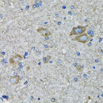
IHC-P analysis of mouse spinal cord tissue using GTX55806 SSBP1 antibody. Dilution : 1:100


IHC-P analysis of mouse spinal cord tissue using GTX55806 SSBP1 antibody. Dilution : 1:100
SSBP1 antibody
GTX55806
ApplicationsImmunoFluorescence, ImmunoPrecipitation, Western Blot, ImmunoCytoChemistry, ImmunoHistoChemistry, ImmunoHistoChemistry Paraffin
Product group Antibodies
ReactivityHuman, Mouse
TargetSSBP1
Overview
- SupplierGeneTex
- Product NameSSBP1 antibody
- Delivery Days Customer9
- Application Supplier NoteWB: 1:500 - 1:2000. ICC/IF: 1:50 - 1:100. IHC-P: 1:50 - 1:200. IP: 1:50 - 1:200. *Optimal dilutions/concentrations should be determined by the researcher.Not tested in other applications.
- ApplicationsImmunoFluorescence, ImmunoPrecipitation, Western Blot, ImmunoCytoChemistry, ImmunoHistoChemistry, ImmunoHistoChemistry Paraffin
- CertificationResearch Use Only
- ClonalityPolyclonal
- ConjugateUnconjugated
- Gene ID6742
- Target nameSSBP1
- Target descriptionsingle stranded DNA binding protein 1
- Target synonymsMt-SSB, OPA13, SOSS-B1, SSBP, mtSSB, single-stranded DNA-binding protein, mitochondrial, PWP1-interacting protein 17, single-stranded DNA binding protein 1, mitochondrial
- HostRabbit
- IsotypeIgG
- Protein IDQ04837
- Protein NameSingle-stranded DNA-binding protein, mitochondrial
- Scientific DescriptionSSBP1 is a housekeeping gene involved in mitochondrial biogenesis (Tiranti et al., 1995 [PubMed 7789991]). It is also a subunit of a single-stranded DNA (ssDNA)-binding complex involved in the maintenance of genome stability (Huang et al., 2009) [PubMed 19683501].[supplied by OMIM, Feb 2010]
- ReactivityHuman, Mouse
- Storage Instruction-20°C or -80°C,2°C to 8°C
- UNSPSC41116161
References
- SSBP1 drives high fructose-induced glomerular podocyte ferroptosis via activating DNA-PK/p53 pathway. Wu WY et al., 2022 Jun, Redox BiolRead this paper
Datasheet
Related products
Product group Antibodies
Anti-MtSSB AntibodyA95215
ApplicationsImmunoFluorescence, Western Blot, ELISA, ImmunoHistoChemistry
ReactivityHuman, Mouse, Rat
- SizePrice

Product group Antibodies
Anti-SSBP1 Antibody Picoband(r)A05166-1-CARRIER-FREE
ApplicationsFlow Cytometry, ImmunoFluorescence, Western Blot, ELISA, ImmunoCytoChemistry, ImmunoHistoChemistry
ReactivityHuman
TargetSSBP1
- SizePrice
Product group Antibodies
SSBP1 AntibodyCSB-PA004162
ApplicationsImmunoFluorescence, Western Blot, ELISA, ImmunoHistoChemistry
ReactivityHuman, Mouse, Rat
TargetSSBP1
- SizePrice
Product group Antibodies

SSBP1 / mtSSB AntibodyLS-C405358
ApplicationsWestern Blot, ELISA, ImmunoHistoChemistry
ReactivityHuman, Mouse, Rat
TargetSSBP1
- SizePrice

Product group Antibodies
Anti-SSBP1 AntibodyHPA002866
ApplicationsWestern Blot, ImmunoCytoChemistry, ImmunoHistoChemistry
ReactivityHuman, Mouse, Rat
TargetSSBP1
- SizePrice