

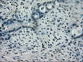
IHC-P analysis of colon adenocarcinoma tissue using GTX83550 STAT1 antibody [15H3]. Antigen retrieval : Heat-induced epitope retrieval by 10mM citrate buffer, pH6.0, 100oC for 10min. Dilution : 1:50
![IHC-P analysis of bladder carcinoma tissue using GTX83550 STAT1 antibody [15H3]. Antigen retrieval : Heat-induced epitope retrieval by 10mM citrate buffer, pH6.0, 100oC for 10min. Dilution : 1:50](https://www.genetex.com/upload/website/prouct_img/normal/GTX83550/GTX83550_1575_IHC-P_w_23061420_601.webp "IHC-P analysis of bladder carcinoma tissue using GTX83550 STAT1 antibody [15H3]. Antigen retrieval : Heat-induced epitope retrieval by 10mM citrate buffer, pH6.0, 100oC for 10min. Dilution : 1:50")
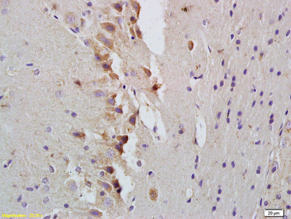
![IHC-P analysis of pancreas carcinoma tissue using GTX83550 STAT1 antibody [15H3]. Antigen retrieval : Heat-induced epitope retrieval by 10mM citrate buffer, pH6.0, 100oC for 10min. Dilution : 1:50](https://www.genetex.com/upload/website/prouct_img/normal/GTX83550/GTX83550_1576_IHC-P_w_23061419_983.webp "IHC-P analysis of pancreas carcinoma tissue using GTX83550 STAT1 antibody [15H3]. Antigen retrieval : Heat-induced epitope retrieval by 10mM citrate buffer, pH6.0, 100oC for 10min. Dilution : 1:50")
![IHC-P analysis of thyroid carcinoma tissue using GTX83550 STAT1 antibody [15H3]. Antigen retrieval : Heat-induced epitope retrieval by 10mM citrate buffer, pH6.0, 100oC for 10min. Dilution : 1:50](https://www.genetex.com/upload/website/prouct_img/normal/GTX83550/GTX83550_1577_IHC-P_w_23061420_758.webp "IHC-P analysis of thyroid carcinoma tissue using GTX83550 STAT1 antibody [15H3]. Antigen retrieval : Heat-induced epitope retrieval by 10mM citrate buffer, pH6.0, 100oC for 10min. Dilution : 1:50")
![WB analysis of HEK293T cells transfected with STAT1 plasmid (Right) or empty vector (Left) for 48 hrs using GTX83550 STAT1 antibody [15H3]. Loading : 5 ug per lane](https://www.genetex.com/upload/website/prouct_img/normal/GTX83550/GTX83550_3722_WB_w_23061420_444.webp "WB analysis of HEK293T cells transfected with STAT1 plasmid (Right) or empty vector (Left) for 48 hrs using GTX83550 STAT1 antibody [15H3]. Loading : 5 ug per lane")
![ICC/IF analysis of COS7 cells transiently transfected with STAT1 plasmid using GTX83550 STAT1 antibody [15H3].](https://www.genetex.com/upload/website/prouct_img/normal/GTX83550/GTX83550_708_ICCIF_w_23061420_418.webp "ICC/IF analysis of COS7 cells transiently transfected with STAT1 plasmid using GTX83550 STAT1 antibody [15H3].")
IHC-P analysis of colon adenocarcinoma tissue using GTX83550 STAT1 antibody [15H3]. Antigen retrieval : Heat-induced epitope retrieval by 10mM citrate buffer, pH6.0, 100oC for 10min. Dilution : 1:50
STAT1 antibody [15H3]
GTX83550
ApplicationsImmunoFluorescence, Western Blot, ImmunoCytoChemistry, ImmunoHistoChemistry, ImmunoHistoChemistry Paraffin
Product group Antibodies
ReactivityHuman
TargetSTAT1
Overview
- SupplierGeneTex
- Product NameSTAT1 antibody [15H3]
- Delivery Days Customer9
- Application Supplier NoteWB: 1:2000. *Optimal dilutions/concentrations should be determined by the researcher.Not tested in other applications.
- ApplicationsImmunoFluorescence, Western Blot, ImmunoCytoChemistry, ImmunoHistoChemistry, ImmunoHistoChemistry Paraffin
- CertificationResearch Use Only
- ClonalityMonoclonal
- Clone ID15H3
- Concentration0.6 mg/ml
- ConjugateUnconjugated
- Gene ID6772
- Target nameSTAT1
- Target descriptionsignal transducer and activator of transcription 1
- Target synonymsCANDF7, IMD31A, IMD31B, IMD31C, ISGF-3, STAT91, signal transducer and activator of transcription 1-alpha/beta, signal transducer and activator of transcription 1, 91kD, signal transducer and activator of transcription 1, 91kDa, transcription factor ISGF-3 components p91/p84
- HostMouse
- IsotypeIgG1
- Protein IDP42224
- Protein NameSignal transducer and activator of transcription 1-alpha/beta
- Scientific DescriptionSignal transducer and activator of transcription that mediates signaling by interferons (IFNs). Following type I IFN (IFN-alpha and IFN-beta) binding to cell surface receptors, Jak kinases (TYK2 and JAK1) are activated, leading to tyrosine phosphorylation of STAT1 and STAT2. The phosphorylated STATs dimerize, associate with ISGF3G/IRF-9 to form a complex termed ISGF3 transcription factor, that enters the nucleus. ISGF3 binds to the IFN stimulated response element (ISRE) to activate the transcription of interferon stimulated genes, which drive the cell in an antiviral state. In response to type II IFN (IFN-gamma), STAT1 is tyrosine- and serine-phosphorylated. It then forms a homodimer termed IFN-gamma-activated factor (GAF), migrates into the nucleus and binds to the IFN gamma activated sequence (GAS) to drive the expression of the target genes, inducing a cellular antiviral state.
- ReactivityHuman
- Storage Instruction-20°C or -80°C,2°C to 8°C
- UNSPSC12352203
Datasheet
Related products
Product group Antibodies
STAT1 Polyclonal AntibodyCAC15929
ApplicationsImmunoPrecipitation, Western Blot, ImmunoHistoChemistry
ReactivityMouse, Primate, Rat
TargetSTAT1
- SizePrice
Product group Antibodies
Anti-P-STAT1 Antibody130-10608
ApplicationsELISA
ReactivityHuman
TargetSTAT1
- SizePrice
Product group Antibodies
References
STAT1 Polyclonal AntibodyBS-1317R
ApplicationsImmunoFluorescence, Western Blot, ELISA, ImmunoCytoChemistry, ImmunoHistoChemistry, ImmunoHistoChemistry Frozen, ImmunoHistoChemistry Paraffin
ReactivityBovine, Canine, Equine, Human, Mouse, Porcine, Rat, Sheep
TargetSTAT1
- SizePrice
Product group Antibodies
Anti-STAT1 AntibodyA100762
ApplicationsWestern Blot, ELISA
ReactivityHuman
- SizePrice
Product group Antibodies

Anti-STAT1 [AbAb1-STAT1]AB04098-10.0
ApplicationsELISA
ReactivityHuman
TargetSTAT1
- SizePrice
![WB analysis of HeLa cell extracts with/without TNF-alpha (20 ng/ml, 37oC, 30 min) treatment using GTX00959 STAT1 (phospho Ser701) antibody [GT1197]. Dilution : 1:1000 Loading : 25microg](https://www.genetex.com/upload/website/prouct_img/normal/GTX00959/GTX00959_20200327_WB_1_w_23053121_463.webp)
Product group Antibodies
ApplicationsWestern Blot
ReactivityHuman, Mouse, Rat
TargetSTAT1
- SizePrice
![FACS analysis of MCF-7 cells using GTX01142 STAT1 antibody [SD20-75]. Red : primary antibody Black : unlabelled control Dilution : 1:50](https://www.genetex.com/upload/website/prouct_img/normal/GTX01142/GTX01142_20200303_FACS_569_w_23053121_212.webp)
Product group Antibodies
STAT1 antibody [SD20-75]GTX01142
ApplicationsFlow Cytometry, Western Blot
ReactivityHuman
TargetSTAT1
- SizePrice
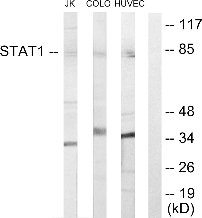
![WB analysis of various samples using GTX01292 STAT1 antibody [GT1157]. Dilution : 1:1000 Loading : 25 microg](https://www.genetex.com/upload/website/prouct_img/normal/GTX01292/GTX01292_20200508_WB_1_w_23053121_586.webp)
Product group Antibodies
STAT1 antibody [GT1157]GTX01292
ApplicationsImmunoPrecipitation, Western Blot
ReactivityHuman, Mouse, Rat
TargetSTAT1
- SizePrice