

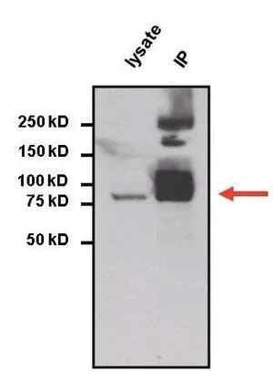
IP analysis of HepG2 cell lysates using GTX15789 STAT3 antibody [9D8]. IP reaction : 2microg antibody / 750microg lysate Dilution : 1:5000
![WB analysis of STAT3 siRNA transfected or non-targeting control transfected U2OS cell lysate using GTX15789 STAT3 antibody [9D8]. Dilution : 1:1000 Loading : 25 microg](https://www.genetex.com/upload/website/prouct_img/normal/GTX15789/GTX15789_1538_WB_w_23060620_817.webp "WB analysis of STAT3 siRNA transfected or non-targeting control transfected U2OS cell lysate using GTX15789 STAT3 antibody [9D8]. Dilution : 1:1000 Loading : 25 microg")
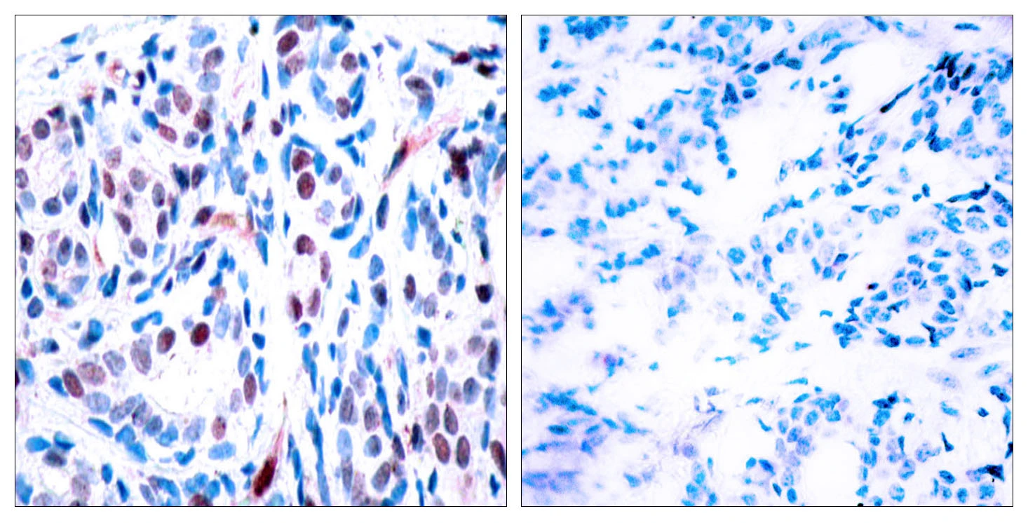
![IHC-P analysis of biopsies of normal and cancer tissues using GTX15789 STAT3 antibody [9D8]. Antigen retrieval : heat induced antigen retrieval was performed using 10mM sodium citrate (pH6.0) buffer for 20 minutes at 95oC Dilution : 1:1600](https://www.genetex.com/upload/website/prouct_img/normal/GTX15789/GTX15789_2042_IHC-P_w_23060620_756.webp "IHC-P analysis of biopsies of normal and cancer tissues using GTX15789 STAT3 antibody [9D8]. Antigen retrieval : heat induced antigen retrieval was performed using 10mM sodium citrate (pH6.0) buffer for 20 minutes at 95oC Dilution : 1:1600")
![ICC/IF analysis of HeLa cells using GTX15789 STAT3 antibody [9D8]. Green : Primary antibody Blue : Nuclei Fixation : Formalin Permeabilization : 0.1% Triton X-100 in TBS for 10 minutes Dilution : 1:100 for at least 1 hour at room temperature](https://www.genetex.com/upload/website/prouct_img/normal/GTX15789/GTX15789_317_ICC-IF_w_23060620_106.webp "ICC/IF analysis of HeLa cells using GTX15789 STAT3 antibody [9D8]. Green : Primary antibody Blue : Nuclei Fixation : Formalin Permeabilization : 0.1% Triton X-100 in TBS for 10 minutes Dilution : 1:100 for at least 1 hour at room temperature")
![ChIP analysis of 1x10? HTC-IR rat hepatoma cells treated with insulin for 0, 10, and 30 minutes using GTX15789 STAT3 antibody [9D8]. Immunoprecipitation was performed using a multiplex microplate Matrix ChIP assay (refer to PMID: 22098709). The precipitated DNA was detected by PCR with primer set targeting to exon-1 or exon-28 of the LAMC1 gene. IP reaction : 1.0ul antibody / 100ul sample](https://www.genetex.com/upload/website/prouct_img/normal/GTX15789/GTX15789_6_ChIP_w_23060620_500.webp "ChIP analysis of 1x10? HTC-IR rat hepatoma cells treated with insulin for 0, 10, and 30 minutes using GTX15789 STAT3 antibody [9D8]. Immunoprecipitation was performed using a multiplex microplate Matrix ChIP assay (refer to PMID: 22098709). The precipitated DNA was detected by PCR with primer set targeting to exon-1 or exon-28 of the LAMC1 gene. IP reaction : 1.0ul antibody / 100ul sample")
IP analysis of HepG2 cell lysates using GTX15789 STAT3 antibody [9D8]. IP reaction : 2microg antibody / 750microg lysate Dilution : 1:5000
STAT3 antibody [9D8]
GTX15789
ApplicationsImmunoFluorescence, ImmunoPrecipitation, Western Blot, ChIP Chromatin ImmunoPrecipitation, ImmunoCytoChemistry, ImmunoHistoChemistry, ImmunoHistoChemistry Paraffin
Product group Antibodies
TargetSTAT3
Overview
- SupplierGeneTex
- Product NameSTAT3 antibody [9D8] - KO/KD, Orthogonal Validated
- Delivery Days Customer9
- Application Supplier NoteWB: 1:5000. ICC/IF: 1:100. IHC-P: 1:1600. IP: 2 microg. ChIP assay: 1-3 microl. *Optimal dilutions/concentrations should be determined by the researcher.Not tested in other applications.
- ApplicationsImmunoFluorescence, ImmunoPrecipitation, Western Blot, ChIP Chromatin ImmunoPrecipitation, ImmunoCytoChemistry, ImmunoHistoChemistry, ImmunoHistoChemistry Paraffin
- CertificationResearch Use Only
- ClonalityMonoclonal
- Clone ID9D8
- Concentration1 mg/ml
- ConjugateUnconjugated
- Gene ID6774
- Target nameSTAT3
- Target descriptionsignal transducer and activator of transcription 3
- Target synonymsacute-phase response factor; ADMIO; ADMIO1; APRF; DNA-binding protein APRF; HIES; signal transducer and activator of transcription 3
- HostMouse
- IsotypeIgG
- Protein IDP40763
- Protein NameSignal transducer and activator of transcription 3
- Scientific DescriptionThe protein encoded by this gene is a member of the STAT protein family. In response to cytokines and growth factors, STAT family members are phosphorylated by the receptor associated kinases, and then form homo- or heterodimers that translocate to the cell nucleus where they act as transcription activators. This protein is activated through phosphorylation in response to various cytokines and growth factors including IFNs, EGF, IL5, IL6, HGF, LIF and BMP2. This protein mediates the expression of a variety of genes in response to cell stimuli, and thus plays a key role in many cellular processes such as cell growth and apoptosis. The small GTPase Rac1 has been shown to bind and regulate the activity of this protein. PIAS3 protein is a specific inhibitor of this protein. Mutations in this gene are associated with infantile-onset multisystem autoimmune disease and hyper-immunoglobulin E syndrome. Alternative splicing results in multiple transcript variants encoding distinct isoforms. [provided by RefSeq, Sep 2015]
- Storage Instruction-20°C or -80°C,2°C to 8°C
- UNSPSC12352203
References
- Mitochondrial Respiratory Defect Enhances Hepatoma Cell Invasiveness via STAT3/NFE2L1/STX12 Axis. Lee YK et al., 2020 Sep 15, Cancers (Basel)Read more
- Targeted regulationof STAT3 by miR-29a in mediating Taxol resistance of nasopharyngeal carcinoma cell line CNE-1. Gao J et al., 2018, Cancer BiomarkRead more
Datasheet
Related products
Product group Antibodies
ApplicationsImmunoPrecipitation
TargetSTAT3
- SizePrice
![ICC/IF analysis of HeLa cells using GTX82752 STAT3 antibody [3B5]. Green : STAT3 Blue: DRAQ5 fluorescent DNA dye Red: Actin filaments](https://www.genetex.com/upload/website/prouct_img/normal/GTX82752/GTX82752_20170912_ICCIF_w_23061322_468.webp)
Product group Antibodies
STAT3 antibody [3B5]GTX82752
ApplicationsImmunoFluorescence, Western Blot, ELISA, ImmunoCytoChemistry, ImmunoHistoChemistry, ImmunoHistoChemistry Paraffin
TargetSTAT3
- SizePrice
![IHC-P analysis of human Liver tissue using GTX83103 STAT3 antibody [7G3H4].](https://www.genetex.com/upload/website/prouct_img/normal/GTX83103/GTX83103_20170912_IHC-P_w_23061322_613.webp)
Product group Antibodies
STAT3 antibody [7G3H4]GTX83103
ApplicationsWestern Blot, ELISA, ImmunoHistoChemistry, ImmunoHistoChemistry Paraffin
TargetSTAT3
- SizePrice
Product group Antibodies
STAT3 (phospho Tyr705) antibodyGTX86741
ApplicationsWestern Blot, ImmunoHistoChemistry, ImmunoHistoChemistry Paraffin
TargetSTAT3
- SizePrice
Product group Antibodies
References
STAT3 antibody [C3], C-termGTX104616
ApplicationsImmunoFluorescence, Western Blot, ImmunoCytoChemistry, ImmunoHistoChemistry, ImmunoHistoChemistry Paraffin
TargetSTAT3
- SizePrice
Product group Antibodies
References
STAT3 (phospho Tyr705) antibodyGTX133464
ApplicationsWestern Blot
TargetSTAT3
- SizePrice
![IHC-P analysis of human renal cell carcinoma tissue using GTX18120 STAT3 antibody [STAT3/2409].](https://www.genetex.com/upload/website/prouct_img/normal/GTX18120/GTX18120_20200115_IHC-P_1086_w_23060620_635.webp)
Product group Antibodies
STAT3 antibody [STAT3/2409]GTX18120
ApplicationsImmunoHistoChemistry, ImmunoHistoChemistry Paraffin, Other Application
TargetSTAT3
- SizePrice
![WB analysis of NIH-3T3 cell extracts with/without TNF-alpha (20 ng/ml, 37oC, 30 min) treatment using GTX00966 STAT3 (phospho Tyr705) antibody [GT1204]. Dilution : 1:1000 Loading : 25microg](https://www.genetex.com/upload/website/prouct_img/normal/GTX00966/GTX00966_20200327_WB_1_w_23053121_254.webp)
Product group Antibodies
ApplicationsWestern Blot
TargetSTAT3
- SizePrice
![IHC-P analysis of rat spleen tissue section using GTX00968 STAT3 (phospho Ser727) antibody [GT1206]. Dilution : 1:100](https://www.genetex.com/upload/website/prouct_img/normal/GTX00968/GTX00968_20200327_IHC-P_32_w_23053121_108.webp)
Product group Antibodies
ApplicationsWestern Blot, ImmunoHistoChemistry, ImmunoHistoChemistry Paraffin
TargetSTAT3
- SizePrice
![WB analysis of Wild-type (WT) and STAT3 knockout (KO) HeLa cell extracts using GTX01294 STAT3 antibody [GT1159]. Dilution : 1:1000 Loading : 25microg](https://www.genetex.com/upload/website/prouct_img/normal/GTX01294/GTX01294_20200327_WB_1_w_23053121_397.webp)