

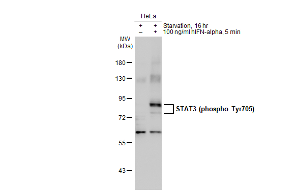
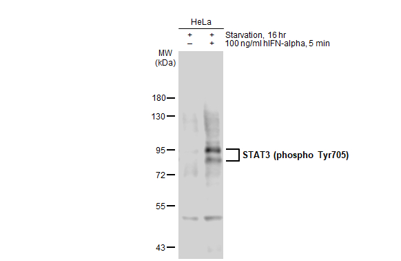
Untreated (–) and treated (+) HeLa whole cell extracts (30 μg) were separated by 7.5% SDS-PAGE, and the membrane was blotted with STAT3 (phospho Tyr705) antibody (GTX133464) diluted at 1:1000. The HRP-conjugated anti-rabbit IgG antibody (GTX213110-01) was used to detect the primary antibody.

Untreated (–) and treated (+) HeLa whole cell extracts (30 μg) were separated by 7.5% SDS-PAGE, and the membrane was blotted with STAT3 (phospho Tyr705) antibody (GTX133464) diluted at 1:1000. The HRP-conjugated anti-rabbit IgG antibody (GTX213110-01) was used to detect the primary antibody.
STAT3 (phospho Tyr705) antibody
GTX133464
ApplicationsWestern Blot
Product group Antibodies
ReactivityHuman, Mouse
TargetSTAT3
Overview
- SupplierGeneTex
- Product NameSTAT3 (phospho Tyr705) antibody
- Delivery Days Customer9
- Application Supplier NoteWB: 1:500-1:3000. *Optimal dilutions/concentrations should be determined by the researcher.Not tested in other applications.
- ApplicationsWestern Blot
- CertificationResearch Use Only
- ClonalityPolyclonal
- Concentration0.66 mg/ml
- ConjugateUnconjugated
- Gene ID6774
- Target nameSTAT3
- Target descriptionsignal transducer and activator of transcription 3
- Target synonymsADMIO, ADMIO1, APRF, HIES, signal transducer and activator of transcription 3, DNA-binding protein APRF, acute-phase response factor
- HostRabbit
- IsotypeIgG
- Protein IDP40763
- Protein NameSignal transducer and activator of transcription 3
- Scientific DescriptionThe protein encoded by this gene is a member of the STAT protein family. In response to cytokines and growth factors, STAT family members are phosphorylated by the receptor associated kinases, and then form homo- or heterodimers that translocate to the cell nucleus where they act as transcription activators. This protein is activated through phosphorylation in response to various cytokines and growth factors including IFNs, EGF, IL5, IL6, HGF, LIF and BMP2. This protein mediates the expression of a variety of genes in response to cell stimuli, and thus plays a key role in many cellular processes such as cell growth and apoptosis. The small GTPase Rac1 has been shown to bind and regulate the activity of this protein. PIAS3 protein is a specific inhibitor of this protein. Three alternatively spliced transcript variants encoding distinct isoforms have been described. [provided by RefSeq]
- ReactivityHuman, Mouse
- Storage Instruction-20°C or -80°C,2°C to 8°C
- UNSPSC41116161
Datasheet
Related products
Product group Antibodies
Anti-STAT3 AntibodyA95775
ApplicationsWestern Blot, ELISA, ImmunoHistoChemistry
ReactivityHuman, Mouse, Rat
- SizePrice
Product group Antibodies
Anti-P-STAT3 Antibody130-10609
ApplicationsELISA
ReactivityHuman
TargetSTAT3
- SizePrice
Product group Antibodies

Anti-STAT3 phosphorylated [AbAb1-pSTAT3]AB04100-10.0
ApplicationsImmunoPrecipitation, Western Blot, ELISA
ReactivityHuman, Mouse
TargetSTAT3
- SizePrice

Product group Antibodies
Anti-STAT3 AntibodyAMAB90776
ApplicationsWestern Blot, ImmunoHistoChemistry
ReactivityHuman
TargetSTAT3
- SizePrice
Product group Antibodies
References
STAT3 Polyclonal AntibodyBS-1141R
ApplicationsFlow Cytometry, ImmunoFluorescence, Western Blot, ELISA, ImmunoCytoChemistry, ImmunoHistoChemistry, ImmunoHistoChemistry Frozen, ImmunoHistoChemistry Paraffin
ReactivityBovine, Canine, Chicken, Guinea Pig, Human, Mouse, Porcine, Rabbit, Rat, Sheep
TargetSTAT3
- SizePrice
Product group Antibodies
STAT3 AntibodyCSB-PA004173
ApplicationsWestern Blot, ELISA, ImmunoHistoChemistry
ReactivityHuman, Mouse, Rat
TargetSTAT3
- SizePrice
Product group Antibodies

ApplicationsFlow Cytometry, ImmunoFluorescence, ELISA, ImmunoHistoChemistry
ReactivityBovine, Human, Mouse, Rat
TargetSTAT3
- SizePrice
Product group Antibodies
Stat3 Polyclonal AntibodyCAC08876
ApplicationsImmunoFluorescence, ImmunoPrecipitation, ELISA, ImmunoHistoChemistry
TargetSTAT3
- SizePrice
Product group Antibodies
STAT3 (phospho Tyr705) antibodyGTX133615
ApplicationsWestern Blot
ReactivityHuman
TargetSTAT3
- SizePrice