

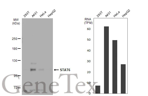
Various whole cell extracts (30 μg) were separated by 5% SDS-PAGE, and the membrane was blotted with STAT6 antibody (GTX113273) diluted at 1:1000. The HRP-conjugated anti-rabbit IgG antibody (GTX213110-01) was used to detect the primary antibody. Corresponding RNA expression data for the same cell lines are based on Human Protein Atlas program.
 diluted at 1:500. Blue: Hoechst 33342 staining.")
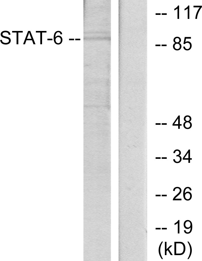
 10% SDS-PAGE The immunoprecipitated STAT6 protein was detected by STAT6 antibody (GTX113273) diluted at 1:1000. EasyBlot anti-rabbit IgG (GTX221666-01) was used as a secondary reagent.")
Various whole cell extracts (30 μg) were separated by 5% SDS-PAGE, and the membrane was blotted with STAT6 antibody (GTX113273) diluted at 1:1000. The HRP-conjugated anti-rabbit IgG antibody (GTX213110-01) was used to detect the primary antibody. Corresponding RNA expression data for the same cell lines are based on Human Protein Atlas program.
STAT6 antibody
GTX113273
ApplicationsImmunoFluorescence, ImmunoPrecipitation, Western Blot, ImmunoCytoChemistry, ImmunoHistoChemistry, ImmunoHistoChemistry Paraffin
Product group Antibodies
ReactivityHuman, Mouse
TargetSTAT6
Overview
- SupplierGeneTex
- Product NameSTAT6 antibody
- Delivery Days Customer9
- Application Supplier NoteWB: 1:500-1:3000. IHC-P: 1:100-1:1000. IP: 1:500-1:1000. *Optimal dilutions/concentrations should be determined by the researcher.Not tested in other applications.
- ApplicationsImmunoFluorescence, ImmunoPrecipitation, Western Blot, ImmunoCytoChemistry, ImmunoHistoChemistry, ImmunoHistoChemistry Paraffin
- CertificationResearch Use Only
- ClonalityPolyclonal
- Concentration0.22 mg/ml
- ConjugateUnconjugated
- Gene ID6778
- Target nameSTAT6
- Target descriptionsignal transducer and activator of transcription 6
- Target synonymsD12S1644, HIES6, IL-4-STAT, STAT6B, STAT6C, signal transducer and activator of transcription 6, STAT, interleukin4-induced, signal transducer and activator of transcription 6, interleukin-4 induced, transcription factor IL-4 STAT
- HostRabbit
- IsotypeIgG
- Protein IDP42226
- Protein NameSignal transducer and activator of transcription 6
- Scientific DescriptionThe protein encoded by this gene is a member of the STAT family of transcription factors. In response to cytokines and growth factors, STAT family members are phosphorylated by the receptor associated kinases, and then form homo- or heterodimers that translocate to the cell nucleus where they act as transcription activators. This protein plays a central role in exerting IL4 mediated biological responses. It is found to induce the expression of BCL2L1/BCL-X(L), which is responsible for the anti-apoptotic activity of IL4. Knockout studies in mice suggested the roles of this gene in differentiation of T helper 2 (Th2) cells, expression of cell surface markers, and class switch of immunoglobulins. [provided by RefSeq]
- ReactivityHuman, Mouse
- Storage Instruction-20°C or -80°C,2°C to 8°C
- UNSPSC41116161
Datasheet
Related products
Product group Antibodies
Anti-STAT6 AntibodyA95206
ApplicationsImmunoPrecipitation, Western Blot, ELISA, ImmunoHistoChemistry
ReactivityHuman, Mouse, Rat
- SizePrice
Product group Antibodies
Anti-STAT6 Antibody144-61646
ApplicationsWestern Blot
ReactivityHuman, Mouse, Rat
TargetSTAT6
- SizePrice
Product group Antibodies

STAT6 Antibody (Tyr641)LS-C769550
ApplicationsWestern Blot, ELISA, ImmunoHistoChemistry, ImmunoHistoChemistry Paraffin
ReactivityHuman, Mouse, Rat
TargetSTAT6
- SizePrice

Product group Antibodies
Anti-STAT6 Antibody Picoband(r)A00523-3-CARRIER-FREE
ApplicationsFlow Cytometry, ImmunoFluorescence, Western Blot, ELISA, ImmunoCytoChemistry, ImmunoHistoChemistry
ReactivityHuman
TargetSTAT6
- SizePrice
Product group Antibodies
STAT6 Polyclonal AntibodyBS-1143R
ApplicationsFlow Cytometry, ImmunoFluorescence, ELISA, ImmunoCytoChemistry, ImmunoHistoChemistry, ImmunoHistoChemistry Frozen, ImmunoHistoChemistry Paraffin
ReactivityCanine, Human, Mouse, Rat
TargetSTAT6
- SizePrice
Product group Antibodies
STAT6 AntibodyCSB-PA004183
ApplicationsImmunoPrecipitation, Western Blot, ELISA, ImmunoHistoChemistry
ReactivityHuman, Mouse, Rat
TargetSTAT6
- SizePrice
Product group Antibodies
ApplicationsImmunoPrecipitation, Western Blot, ImmunoCytoChemistry, ImmunoHistoChemistry
ReactivityMouse
TargetSTAT6
- SizePrice
![IHC-P analysis of human renal cell carcinoma tissue using GTX17816 STAT6 antibody [STAT6/2410].](https://www.genetex.com/upload/website/prouct_img/normal/GTX17816/GTX17816_20200115_IHC-P_1087_w_23060620_981.webp)
Product group Antibodies
STAT6 antibody [STAT6/2410]GTX17816
ApplicationsImmunoHistoChemistry, ImmunoHistoChemistry Paraffin, Other Application
ReactivityHuman
TargetSTAT6
- SizePrice
Product group Antibodies
STAT6 antibodyGTX22424
ApplicationsWestern Blot
ReactivityHuman
TargetSTAT6
- SizePrice