

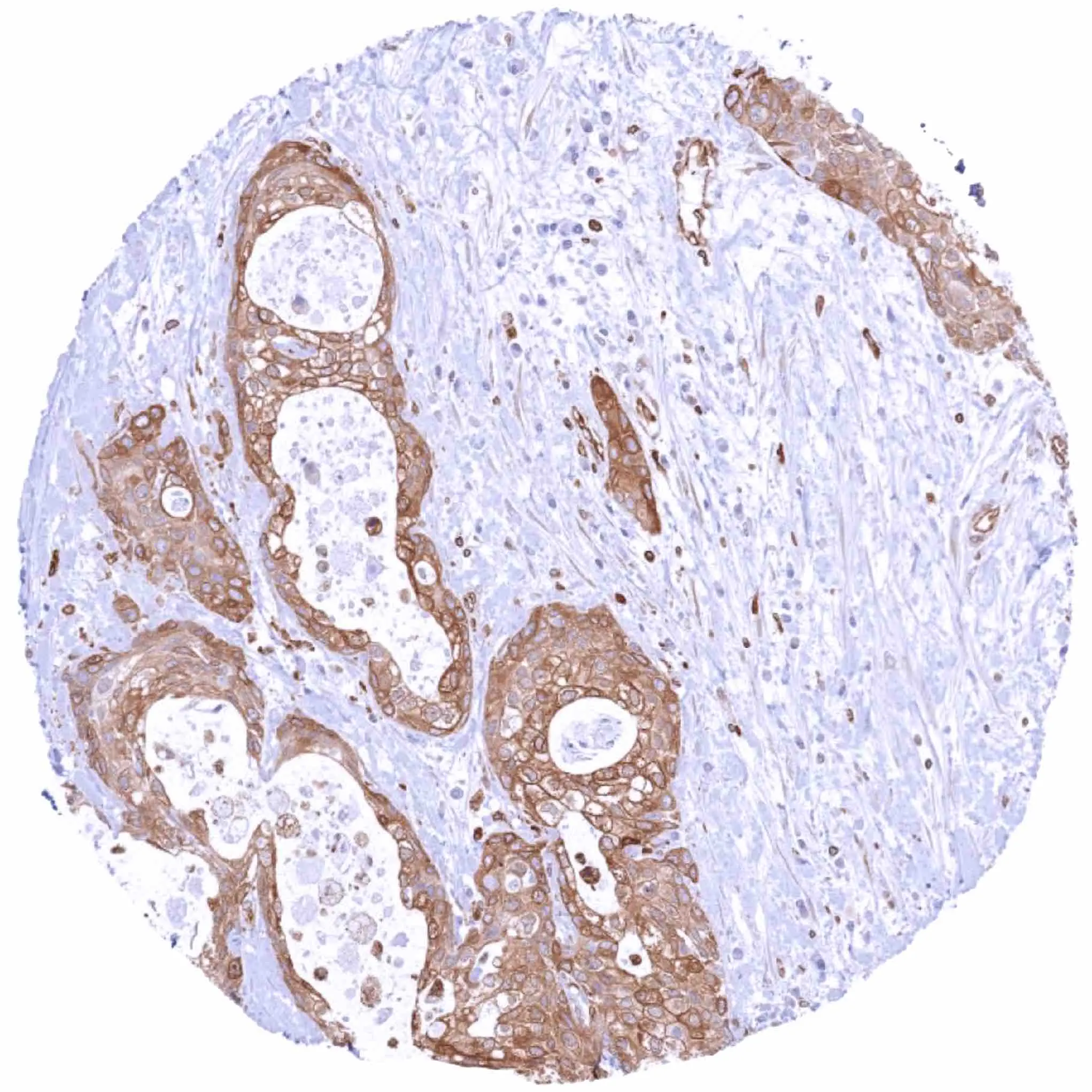
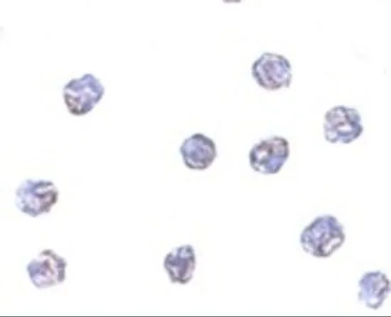
IHC-P analysis of human pancreatic ductal adenocarcinoma (PDAC) tissue using GTX04484 STING antibody [MSVA-515M] HistoMAX?. Ductal adenocarcinoma with strong STING positivity of all tumor cells.
![IHC-P analysis of human adrenal gland tissue using GTX04484 STING antibody [MSVA-515M] HistoMAX?. STING staining of endothelial cells and of macrophages STING immunohistochemistry.](https://www.genetex.com/upload/website/prouct_img/normal/GTX04484/GTX04484_20230728_IHC-P_243_23072722_839.webp "IHC-P analysis of human adrenal gland tissue using GTX04484 STING antibody [MSVA-515M] HistoMAX?. STING staining of endothelial cells and of macrophages STING immunohistochemistry.")
![IHC-P analysis of human mucosa from appendix tissue using GTX04484 STING antibody [MSVA-515M] HistoMAX?. STING staining of inflammatory cells predominates in the interfollicular zone STING immunohistochemistry.](https://www.genetex.com/upload/website/prouct_img/normal/GTX04484/GTX04484_20230728_IHC-P_373_23072723_273.webp "IHC-P analysis of human mucosa from appendix tissue using GTX04484 STING antibody [MSVA-515M] HistoMAX?. STING staining of inflammatory cells predominates in the interfollicular zone STING immunohistochemistry.")
IHC-P analysis of human pancreatic ductal adenocarcinoma (PDAC) tissue using GTX04484 STING antibody [MSVA-515M] HistoMAX?. Ductal adenocarcinoma with strong STING positivity of all tumor cells.
STING antibody [MSVA-515M] HistoMAX(tm)
GTX04484
ApplicationsImmunoHistoChemistry, ImmunoHistoChemistry Paraffin
Product group Antibodies
ReactivityHuman
TargetSTING1
Overview
- SupplierGeneTex
- Product NameSTING antibody [MSVA-515M] HistoMAX(tm)
- Delivery Days Customer9
- Application Supplier NoteIHC-P: 1:100-1:200. *Optimal dilutions/concentrations should be determined by the researcher.Not tested in other applications.
- ApplicationsImmunoHistoChemistry, ImmunoHistoChemistry Paraffin
- CertificationResearch Use Only
- ClonalityMonoclonal
- Clone IDMSVA-515M
- Concentration0.2 mg/ml
- ConjugateUnconjugated
- Gene ID340061
- Target nameSTING1
- Target descriptionstimulator of interferon response cGAMP interactor 1
- Target synonymsERIS, MITA, MPYS, NET23, SAVI, STING, STING-beta, TMEM173, hMITA, hSTING, stimulator of interferon genes protein, N-terminal methionine-proline-tyrosine-serine plasma membrane tetraspanner, endoplasmic reticulum IFN stimulator, endoplasmic reticulum interferon stimulator, mitochondrial mediator of IRF3 activation, stimulator of interferon protein, stimulator of interferon response cGAMP interactor-deltaC, stimulator of interferon response cGAMP interactor-deltaN, sting 1, transmembrane protein 173
- HostMouse
- IsotypeIgG2c
- Protein IDQ86WV6
- Protein NameStimulator of interferon genes protein
- Scientific DescriptionThis gene encodes a five transmembrane protein that functions as a major regulator of the innate immune response to viral and bacterial infections. The encoded protein is a pattern recognition receptor that detects cytosolic nucleic acids and transmits signals that activate type I interferon responses. The encoded protein has also been shown to play a role in apoptotic signaling by associating with type II major histocompatibility complex. Mutations in this gene are the cause of infantile-onset STING-associated vasculopathy. Alternate splicing results in multiple transcript variants. [provided by RefSeq, Sep 2014]
- ReactivityHuman
- Storage Instruction-20°C or -80°C,2°C to 8°C
- UNSPSC41116161
Datasheet
Related products
Product group Antibodies
Anti-TMEM173 AntibodyA40358
ApplicationsWestern Blot, ELISA
ReactivityHuman, Mouse
- SizePrice
Product group Antibodies
Anti-TMEM173 Antibody144-03575
ApplicationsImmunoPrecipitation, Western Blot, ImmunoHistoChemistry
ReactivityHuman, Mouse
TargetSTING1
- SizePrice
Product group Antibodies
STING1 Polyclonal AntibodyBS-8335R
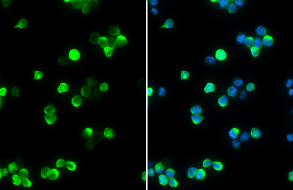
ApplicationsImmunoFluorescence, ELISA, ImmunoCytoChemistry, ImmunoHistoChemistry, ImmunoHistoChemistry Frozen, ImmunoHistoChemistry Paraffin
ReactivityHuman, Mouse, Rat
TargetSTING1
- SizePrice
Product group Antibodies
Tmem173 Polyclonal AntibodyCAC08033
ApplicationsImmunoFluorescence, Western Blot, ELISA, ImmunoHistoChemistry
TargetSTING1
- SizePrice
Product group Antibodies
TMEM173 AntibodyCSB-PA005903
ApplicationsWestern Blot, ELISA
ReactivityHuman, Mouse
TargetSTING1
- SizePrice
Product group Antibodies
STING antibodyGTX134373
ApplicationsImmunoFluorescence, ImmunoPrecipitation, Western Blot, ImmunoCytoChemistry, ImmunoHistoChemistry, ImmunoHistoChemistry Paraffin
ReactivityHuman
TargetSTING1
- SizePrice
Product group Antibodies

TMEM173 / STING AntibodyLS-C332665
ApplicationsWestern Blot, ImmunoHistoChemistry
ReactivityHuman, Mouse
TargetSTING1
- SizePrice
Product group Antibodies
References
STING antibodyGTX85266
ApplicationsImmunoFluorescence, Western Blot, ELISA, ImmunoCytoChemistry
ReactivityHuman, Mouse
TargetSTING1
- SizePrice
Product group Antibodies
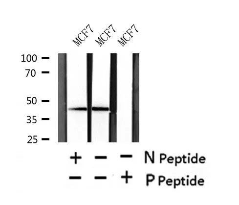
STING (phospho Ser366) antibodyGTX57223
ApplicationsWestern Blot
ReactivityHuman
TargetSTING1
- SizePrice