

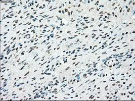
IHC-P analysis of ovary tissue using GTX83546 STK3 antibody [4G10]. Antigen retrieval : Heat-induced epitope retrieval by 10mM citrate buffer, pH6.0, 100oC for 10min. Dilution : 1:50
![IHC-P analysis of endometrium tissue using GTX83546 STK3 antibody [4G10]. Antigen retrieval : Heat-induced epitope retrieval by 10mM citrate buffer, pH6.0, 100oC for 10min. Dilution : 1:50](https://www.genetex.com/upload/website/prouct_img/normal/GTX83546/GTX83546_1545_IHC-P_w_23061420_881.webp "IHC-P analysis of endometrium tissue using GTX83546 STK3 antibody [4G10]. Antigen retrieval : Heat-induced epitope retrieval by 10mM citrate buffer, pH6.0, 100oC for 10min. Dilution : 1:50")
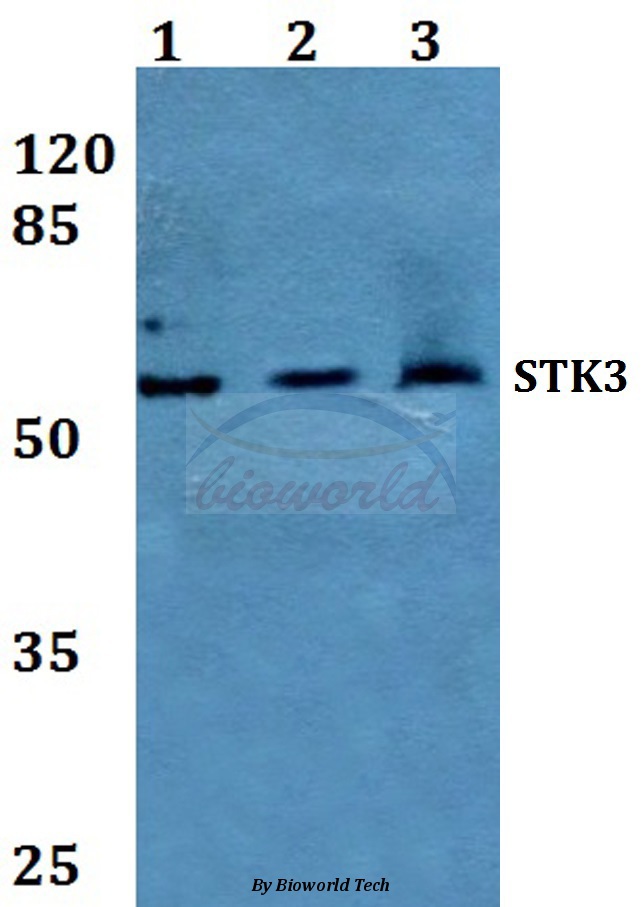
![WB analysis of various samples using GTX83546 STK3 antibody [4G10]. Loading : 10 ug per lane Dilution : 1:200](https://www.genetex.com/upload/website/prouct_img/normal/GTX83546/GTX83546_3429_WB_w_23061420_192.webp "WB analysis of various samples using GTX83546 STK3 antibody [4G10]. Loading : 10 ug per lane Dilution : 1:200")
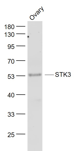
![WB analysis of human tissues (Lane 1-Testis ; Lane 2-Omentum ; Lane 3-Uterus ; Lane 4-Breast ; Lane 5-Brain ; Lane 6-Liver ; Lane 7-Ovary ; Lane 8-Thyroid gland ; Lane 9-colon ; Lane 10-spleen) using GTX83546 STK3 antibody [4G10]. Loading : 10 ug per lane Dilution : 1:200](https://www.genetex.com/upload/website/prouct_img/normal/GTX83546/GTX83546_3656_WB_w_23061420_745.webp "WB analysis of human tissues (Lane 1-Testis ; Lane 2-Omentum ; Lane 3-Uterus ; Lane 4-Breast ; Lane 5-Brain ; Lane 6-Liver ; Lane 7-Ovary ; Lane 8-Thyroid gland ; Lane 9-colon ; Lane 10-spleen) using GTX83546 STK3 antibody [4G10]. Loading : 10 ug per lane Dilution : 1:200")
![IHC-P analysis of lung tissue using GTX83546 STK3 antibody [4G10]. Antigen retrieval : Heat-induced epitope retrieval by 10mM citrate buffer, pH6.0, 100oC for 10min. Dilution : 1:50](https://www.genetex.com/upload/website/prouct_img/normal/GTX83546/GTX83546_1548_IHC-P_w_23061420_861.webp "IHC-P analysis of lung tissue using GTX83546 STK3 antibody [4G10]. Antigen retrieval : Heat-induced epitope retrieval by 10mM citrate buffer, pH6.0, 100oC for 10min. Dilution : 1:50")
![IHC-P analysis of kidney carcinoma tissue using GTX83546 STK3 antibody [4G10]. Antigen retrieval : Heat-induced epitope retrieval by 10mM citrate buffer, pH6.0, 100oC for 10min. Dilution : 1:50](https://www.genetex.com/upload/website/prouct_img/normal/GTX83546/GTX83546_1543_IHC-P_w_23061420_706.webp "IHC-P analysis of kidney carcinoma tissue using GTX83546 STK3 antibody [4G10]. Antigen retrieval : Heat-induced epitope retrieval by 10mM citrate buffer, pH6.0, 100oC for 10min. Dilution : 1:50")
![IHC-P analysis of kidney tissue using GTX83546 STK3 antibody [4G10]. Antigen retrieval : Heat-induced epitope retrieval by 10mM citrate buffer, pH6.0, 100oC for 10min. Dilution : 1:50](https://www.genetex.com/upload/website/prouct_img/normal/GTX83546/GTX83546_1546_IHC-P_w_23061420_748.webp "IHC-P analysis of kidney tissue using GTX83546 STK3 antibody [4G10]. Antigen retrieval : Heat-induced epitope retrieval by 10mM citrate buffer, pH6.0, 100oC for 10min. Dilution : 1:50")
![WB analysis of HEK293T cells transfected with STK3 plasmid (Right) or empty vector (Left) for 48 hrs using GTX83546 STK3 antibody [4G10]. Loading : 5 ug per lane](https://www.genetex.com/upload/website/prouct_img/normal/GTX83546/GTX83546_3712_WB_w_23061420_626.webp "WB analysis of HEK293T cells transfected with STK3 plasmid (Right) or empty vector (Left) for 48 hrs using GTX83546 STK3 antibody [4G10]. Loading : 5 ug per lane")
![WB analysis of various cell lines using GTX83546 STK3 antibody [4G10]. Loading : 35 ug per lane](https://www.genetex.com/upload/website/prouct_img/normal/GTX83546/GTX83546_3714_WB_w_23061420_746.webp "WB analysis of various cell lines using GTX83546 STK3 antibody [4G10]. Loading : 35 ug per lane")
![IHC-P analysis of liver tissue using GTX83546 STK3 antibody [4G10]. Antigen retrieval : Heat-induced epitope retrieval by 10mM citrate buffer, pH6.0, 100oC for 10min. Dilution : 1:50](https://www.genetex.com/upload/website/prouct_img/normal/GTX83546/GTX83546_1547_IHC-P_w_23061420_962.webp "IHC-P analysis of liver tissue using GTX83546 STK3 antibody [4G10]. Antigen retrieval : Heat-induced epitope retrieval by 10mM citrate buffer, pH6.0, 100oC for 10min. Dilution : 1:50")
IHC-P analysis of ovary tissue using GTX83546 STK3 antibody [4G10]. Antigen retrieval : Heat-induced epitope retrieval by 10mM citrate buffer, pH6.0, 100oC for 10min. Dilution : 1:50
STK3 antibody [4G10]
GTX83546
ApplicationsWestern Blot, ImmunoHistoChemistry, ImmunoHistoChemistry Paraffin
Product group Antibodies
ReactivityCanine, Human, Monkey, Mouse
TargetSTK3
Overview
- SupplierGeneTex
- Product NameSTK3 antibody [4G10]
- Delivery Days Customer9
- Application Supplier NoteWB: 1:2000. IHC-P: 1:50. *Optimal dilutions/concentrations should be determined by the researcher.Not tested in other applications.
- ApplicationsWestern Blot, ImmunoHistoChemistry, ImmunoHistoChemistry Paraffin
- CertificationResearch Use Only
- ClonalityMonoclonal
- Clone ID4G10
- Concentration1 mg/ml
- ConjugateUnconjugated
- Gene ID6788
- Target nameSTK3
- Target descriptionserine/threonine kinase 3
- Target synonymsKRS1, MST2, serine/threonine-protein kinase 3, KB-1458E12.1, MST-2, STE20-like kinase MST2, epididymis secretory sperm binding protein, hippo homolog, mammalian STE20-like protein kinase 2, serine/threonine kinase 3 (STE20 homolog, yeast), serine/threonine kinase 3 (Ste20, yeast homolog), serine/threonine-protein kinase Krs-1
- HostMouse
- IsotypeIgG2b
- Protein IDQ13188
- Protein NameSerine/threonine-protein kinase 3
- Scientific DescriptionStress-activated, pro-apoptotic kinase which, following caspase-cleavage, enters the nucleus and induces chromatin condensation followed by internucleosomal DNA fragmentation. Key component of the Hippo signaling pathway which plays a pivotal role in organ size control and tumor suppression by restricting proliferation and promoting apoptosis. The core of this pathway is composed of a kinase cascade wherein MST1/MST2, in complex with its regulatory protein SAV1, phosphorylates and activates LATS1/2 in complex with its regulatory protein MOB1, which in turn phosphorylates and inactivates YAP1 oncoprotein and WWTR1/TAZ. Phosphorylation of YAP1 by LATS2 inhibits its translocation into the nucleus to regulate cellular genes important for cell proliferation, cell death, and cell migration. MST1/MST2 are required to repress proliferation of mature hepatocytes, to prevent activation of facultative adult liver stem cells (oval cells), and to inhibit tumor formation. Phosphorylates NKX2-1.
- ReactivityCanine, Human, Monkey, Mouse
- Storage Instruction-20°C or -80°C,2°C to 8°C
- UNSPSC41116161
Datasheet
Related products
Product group Antibodies
Anti-STK3 AntibodyA28430
ApplicationsWestern Blot
ReactivityHuman, Mouse, Rat
- SizePrice
Product group Antibodies
Anti-STK3 Antibody144-06992
ApplicationsWestern Blot, ImmunoHistoChemistry
ReactivityHuman, Mouse, Rat
TargetSTK3
- SizePrice
Product group Antibodies
MST3/STK3 Polyclonal AntibodyBS-7599R
ApplicationsImmunoFluorescence, Western Blot, ELISA, ImmunoCytoChemistry, ImmunoHistoChemistry, ImmunoHistoChemistry Frozen, ImmunoHistoChemistry Paraffin
ReactivityBovine, Canine, Chicken, Equine, Human, Mouse, Rabbit, Rat
TargetSTK3
- SizePrice

Product group Antibodies
Anti-STK3/MST-2 Antibody Picoband(r)A02224-2-CARRIER-FREE
ApplicationsFlow Cytometry, ImmunoFluorescence, Western Blot, ELISA, ImmunoCytoChemistry, ImmunoHistoChemistry
ReactivityHuman, Monkey
TargetSTK3
- SizePrice
Product group Antibodies

Goat anti-MST2 / STK3EB11363
ApplicationsWestern Blot, ELISA
ReactivityBovine, Canine, Human, Mouse, Porcine, Rat
TargetSTK3
- SizePrice
Product group Antibodies
Stk3 Polyclonal AntibodyCAC10614
ApplicationsImmunoFluorescence, Western Blot, ELISA, ImmunoHistoChemistry
ReactivityMouse
TargetSTK3
- SizePrice
Product group Antibodies
STK3/STK4 AntibodyCSB-PA050173
ApplicationsWestern Blot, ELISA, ImmunoHistoChemistry
ReactivityHuman, Mouse
TargetSTK3
- SizePrice
Product group Antibodies

STK3 AntibodyLS-C346174
ApplicationsImmunoFluorescence, Western Blot, ImmunoHistoChemistry
ReactivityHuman, Mouse, Rat
TargetSTK3
- SizePrice

Product group Antibodies
Anti-STK3 AntibodyHPA007120
ApplicationsWestern Blot, ImmunoCytoChemistry, ImmunoHistoChemistry
ReactivityHuman, Mouse, Rat
TargetSTK3
- SizePrice