


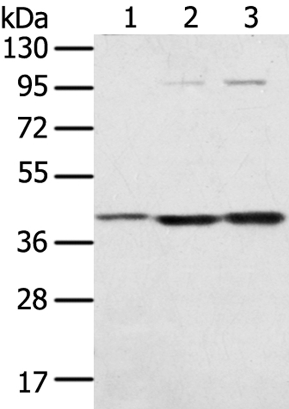
Gel: 8%SDS-PAGE Lysate: 40 microg Lane 1-3: Hepg2 cells TM4 cells Raw264.7 cells Primary antibody: TA350473 (STX18 Antibody) at dilution 1/300 Secondary antibody: Goat anti rabbit IgG at 1/8000 dilution Exposure time: 20 seconds
Gel: 8%SDS-PAGE Lysate: 40 microg Lane 1-3: Hepg2 cells TM4 cells Raw264.7 cells Primary antibody: TA350473 (STX18 Antibody) at dilution 1/300 Secondary antibody: Goat anti rabbit IgG at 1/8000 dilution Exposure time: 20 seconds
Syntaxin 18 (STX18) Rabbit Polyclonal Antibody
TA350473
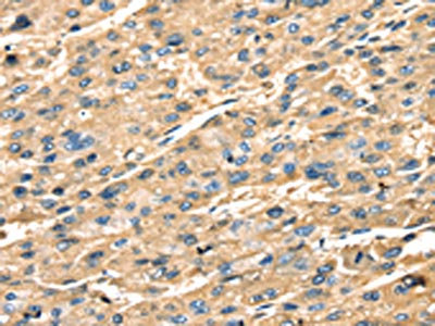
ApplicationsWestern Blot, ImmunoHistoChemistry
Product group Antibodies
ReactivityHuman, Mouse, Rat
TargetSTX18
Overview
- SupplierOriGene
- Product NameSyntaxin 18 (STX18) Rabbit Polyclonal Antibody
- Delivery Days Customer14
- ApplicationsWestern Blot, ImmunoHistoChemistry
- CertificationResearch Use Only
- ClonalityPolyclonal
- Gene ID53407
- Target nameSTX18
- Target descriptionsyntaxin 18
- Target synonymsUfe1, syntaxin-18, cell growth-inhibiting gene 9 protein
- HostRabbit
- IsotypeIgG
- Protein IDQ9P2W9
- Protein NameSyntaxin-18
- Scientific DescriptionRabbit Polyclonal Anti-STX18 rabbit polyclonal antibody
- ReactivityHuman, Mouse, Rat
- Storage Instruction-20°C
- UNSPSC12352203
MSDS
Related products

Product group Antibodies
Anti-Syntaxin 18/STX18 Antibody Picoband(r)A11895-CARRIER-FREE
ApplicationsFlow Cytometry, ImmunoFluorescence, Western Blot, ELISA, ImmunoHistoChemistry
ReactivityHuman
TargetSTX18
- SizePrice
Product group Antibodies
Anti-STX18 AntibodyA38180
ApplicationsWestern Blot, ImmunoHistoChemistry
ReactivityHuman, Mouse
- SizePrice
Product group Antibodies

STX18 / Syntaxin 18 AntibodyLS-C749375
ApplicationsWestern Blot
ReactivityHuman, Mouse, Rat
TargetSTX18
- SizePrice

Product group Antibodies
Anti-STX18 AntibodyHPA003019
ApplicationsImmunoCytoChemistry, ImmunoHistoChemistry
ReactivityHuman
TargetSTX18
- SizePrice
Product group Antibodies
STX18 AntibodyCSB-PA041704
ApplicationsWestern Blot, ELISA, ImmunoHistoChemistry
ReactivityHuman, Mouse, Rat
TargetSTX18
- SizePrice