

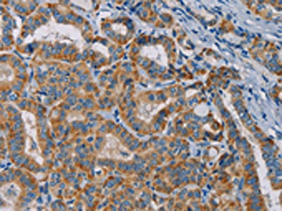
The image is immunohistochemistry of paraffin-embedded Human thyroid cancer tissue using CSB-PA133615(TAB3 Antibody) at dilution 1/25. (Original magnification: x200)
 at dilution 1/25. (Original magnification: x200)")
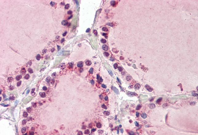
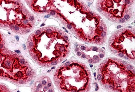
 at dilution 1/800, Secondary antibody: Goat anti rabbit IgG at 1/8000 dilution, Exposure time: 10 seconds")
The image is immunohistochemistry of paraffin-embedded Human thyroid cancer tissue using CSB-PA133615(TAB3 Antibody) at dilution 1/25. (Original magnification: x200)
TAB3 Antibody
CSB-PA133615
ApplicationsWestern Blot, ELISA, ImmunoHistoChemistry
Product group Antibodies
ReactivityHuman, Mouse
TargetTAB3
Overview
- SupplierCusabio
- Product NameTAB3 Antibody
- Delivery Days Customer20
- ApplicationsWestern Blot, ELISA, ImmunoHistoChemistry
- CertificationResearch Use Only
- ClonalityPolyclonal
- ConjugateUnconjugated
- Gene ID257397
- Target nameTAB3
- Target descriptionTGF-beta activated kinase 1 (MAP3K7) binding protein 3
- Target synonymsMAP3K7IP3, NAP1, TGF-beta-activated kinase 1 and MAP3K7-binding protein 3, NF-kappa-B-activating protein 1, NFkB activating protein 1, TAB-3, TAK1-binding protein 3, TGF-beta activated kinase 1 and MAP3K7 binding protein 3, TGF-beta-activated kinase 1-binding protein 3, mitogen-activated protein kinase kinase kinase 7 interacting protein 3
- HostRabbit
- IsotypeIgG
- Protein IDQ8N5C8
- Protein NameTGF-beta-activated kinase 1 and MAP3K7-binding protein 3
- Scientific DescriptionThe product of this gene functions in the NF-kappaB signal transduction pathway. The encoded protein, and the similar and functionally redundant protein MAP3K7IP2/TAB2, forms a ternary complex with the protein kinase MAP3K7/TAK1 and either TRAF2 or TRAF6 in response to stimulation with the pro-inflammatory cytokines TNF or IL-1. Subsequent MAP3K7/TAK1 kinase activity triggers a signaling cascade leading to activation of the NF-kappaB transcription factor. The human genome contains a related pseudogene.
- ReactivityHuman, Mouse
- Storage Instruction-20°C or -80°C
- UNSPSC41116161
Related products
Product group Antibodies
Anti-TAB3 AntibodyA84414
ApplicationsELISA, ImmunoHistoChemistry
ReactivityHuman
- SizePrice

Product group Antibodies
Anti-TAB3 Antibody Picoband(r)A05084-3-CARRIER-FREE
ApplicationsFlow Cytometry, Western Blot, ELISA
ReactivityHuman
TargetTAB3
- SizePrice
Product group Antibodies
Anti-NAP1 / TAB3 (aa59-69) Antibody119-16393
ApplicationsELISA, ImmunoHistoChemistry, ImmunoHistoChemistry Paraffin
ReactivityHuman
TargetTAB3
- SizePrice
Product group Antibodies
MAP3K7IP3 Recombinant Antibody, AbBy Fluor-555 ConjugatedBSM-62098R-BF555
ApplicationsWestern Blot
ReactivityHuman, Mouse
TargetTAB3
- SizePrice
Product group Antibodies

NAP1 / TAB3 AntibodyLS-C401827
ApplicationsWestern Blot, ELISA, ImmunoHistoChemistry
ReactivityHuman, Mouse
TargetTAB3
- SizePrice
Product group Antibodies
TAB3 antibodyGTX31693
ApplicationsELISA, ImmunoHistoChemistry, ImmunoHistoChemistry Paraffin
ReactivityHuman
TargetTAB3
- SizePrice

Product group Antibodies
Anti-TAB3 AntibodyHPA034981
ApplicationsImmunoCytoChemistry, ImmunoHistoChemistry
ReactivityHuman
TargetTAB3
- SizePrice
Product group Antibodies
Anti-TAB3 AntibodyCAB18681
ApplicationsWestern Blot, ELISA, ImmunoHistoChemistry, ImmunoHistoChemistry Paraffin
ReactivityHuman
TargetTAB3
- SizePrice