

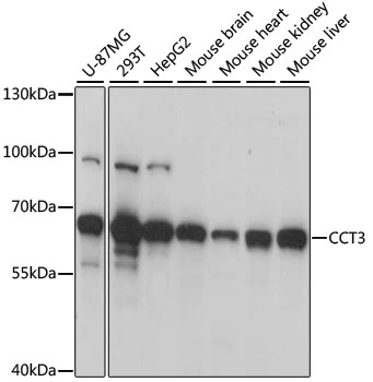
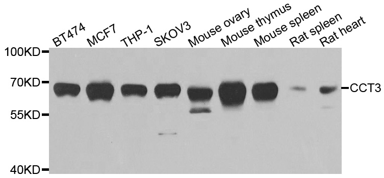
WB analysis of various sample lysates using GTX33073 TCP1 gamma antibody. Dilution : 1:1000 Loading : 25μg per lane


WB analysis of various sample lysates using GTX33073 TCP1 gamma antibody. Dilution : 1:1000 Loading : 25μg per lane
TCP1 gamma antibody
GTX33073
ApplicationsImmunoFluorescence, Western Blot, ImmunoCytoChemistry, ImmunoHistoChemistry, ImmunoHistoChemistry Paraffin
Product group Antibodies
ReactivityHuman, Mouse
TargetCCT3
Overview
- SupplierGeneTex
- Product NameTCP1 gamma antibody
- Delivery Days Customer9
- Application Supplier NoteWB: 1:500 - 1:2000. ICC/IF: 1:50 - 1:200. IHC-P: 1:50 - 1:200. *Optimal dilutions/concentrations should be determined by the researcher.Not tested in other applications.
- ApplicationsImmunoFluorescence, Western Blot, ImmunoCytoChemistry, ImmunoHistoChemistry, ImmunoHistoChemistry Paraffin
- CertificationResearch Use Only
- ClonalityPolyclonal
- ConjugateUnconjugated
- Gene ID7203
- Target nameCCT3
- Target descriptionchaperonin containing TCP1 subunit 3
- Target synonymsCCT-gamma, CCTG, NEDSVH, PIG48, TCP-1-gamma, TRIC5, T-complex protein 1 subunit gamma, T-complex protein 1, gamma subunit, TCP1 (t-complex-1) ring complex, polypeptide 5, chaperonin containing T-complex polypeptide 1 subunit 3, chaperonin containing TCP1, subunit 3 (gamma), hTRiC5
- HostRabbit
- IsotypeIgG
- Protein IDP49368
- Protein NameT-complex protein 1 subunit gamma
- Scientific DescriptionThe protein encoded by this gene is a molecular chaperone that is a member of the chaperonin containing TCP1 complex (CCT), also known as the TCP1 ring complex (TRiC). This complex consists of two identical stacked rings, each containing eight different proteins. Unfolded polypeptides enter the central cavity of the complex and are folded in an ATP-dependent manner. The complex folds various proteins, including actin and tubulin. Alternate transcriptional splice variants have been characterized for this gene. In addition, a pseudogene of this gene has been found on chromosome 8. [provided by RefSeq, Aug 2010]
- ReactivityHuman, Mouse
- Storage Instruction-20°C or -80°C,2°C to 8°C
- UNSPSC41116161
References
- Chaperonin-Containing TCP-1 Promotes Cancer Chemoresistance and Metastasis through the AKT-GSK3beta-beta-Catenin and XIAP-Survivin Pathways. Chang YX et al., 2020 Dec 21, Cancers (Basel)Read this paper
Datasheet
Related products
Product group Antibodies
CCT3 AntibodyCSB-PA004857ESR1HU
ApplicationsWestern Blot, ELISA, ImmunoHistoChemistry
ReactivityHuman, Mouse
TargetCCT3
- SizePrice
Product group Antibodies
Anti-CCT3 AntibodyA30089
ApplicationsImmunoFluorescence, Western Blot, ImmunoHistoChemistry
ReactivityHuman, Mouse, Rat
- SizePrice
Product group Antibodies

Goat anti-CCT3 / TCP1EB09403
ApplicationsWestern Blot, ELISA, ImmunoHistoChemistry
ReactivityBovine, Canine, Human, Mouse, Porcine, Rat
TargetCCT3
- SizePrice

Product group Antibodies
Anti-CCT3 AntibodyHPA006543
ApplicationsWestern Blot, ImmunoCytoChemistry
ReactivityHuman, Mouse, Rat
TargetCCT3
- SizePrice
Product group Antibodies

CCT3 AntibodyLS-C334788
ApplicationsImmunoFluorescence, Western Blot, ImmunoHistoChemistry
ReactivityHuman, Mouse, Rat
TargetCCT3
- SizePrice
Product group Antibodies
TCP1 gamma antibody, C-termGTX88407
ApplicationsWestern Blot, ImmunoHistoChemistry, ImmunoHistoChemistry Paraffin
ReactivityHuman, Mouse
TargetCCT3
- SizePrice

Product group Antibodies
Anti-CCT3 Antibody Picoband(r)PB9926-CARRIER-FREE
ApplicationsFlow Cytometry, ImmunoFluorescence, Western Blot, ImmunoCytoChemistry, ImmunoHistoChemistry
ReactivityHuman, Mouse, Rat
TargetCCT3
- SizePrice
Product group Antibodies
Anti-CCT3 Antibody144-06547
ApplicationsImmunoFluorescence, Western Blot, ImmunoHistoChemistry
ReactivityHuman, Mouse, Rat
TargetCCT3
- SizePrice