

TERF1 antibody
70R-20764
Product group Antibodies
Overview
- SupplierBiosynth
- Product NameTERF1 antibody
- Delivery Days Customer11
- CertificationResearch Use Only
- UNSPSC41116161
Related products
Product group Antibodies
Anti-TERF1 AntibodyA39736
ApplicationsImmunoFluorescence, Western Blot
ReactivityHuman
- SizePrice

Product group Antibodies
Anti-TRF1/TERF1 Antibody Picoband(r)A02474-2-CARRIER-FREE
ApplicationsImmunoFluorescence, Western Blot, ELISA, ImmunoCytoChemistry
ReactivityHuman
TargetTERF1
- SizePrice
Product group Antibodies
Anti-TERF1 Antibody144-60007
ApplicationsWestern Blot, ImmunoHistoChemistry
ReactivityHuman, Mouse
TargetTERF1
- SizePrice
Product group Antibodies

TRF1 / TERF1 AntibodyLS-C830281
ApplicationsWestern Blot, ELISA, ImmunoHistoChemistry
ReactivityHuman, Mouse
TargetTERF1
- SizePrice
Product group Antibodies
TRF1 Recombinant Antibody, Biotin ConjugatedBSM-61416R-BIOTIN
ApplicationsWestern Blot
ReactivityHuman, Mouse
TargetTERF1
- SizePrice
Product group Antibodies

Goat anti-TERF1 / PIN2EB05325
ApplicationsWestern Blot, ELISA
ReactivityBovine, Human, Porcine
TargetTERF1
- SizePrice
Product group Antibodies
ApplicationsImmunoPrecipitation, Western Blot, ImmunoCytoChemistry, ImmunoHistoChemistry
ReactivityRat
TargetTERF1
- SizePrice
Product group Antibodies
TERF1 AntibodyCSB-PA023386EA01HU
ApplicationsELISA, ImmunoHistoChemistry
ReactivityHuman
TargetTERF1
- SizePrice

Product group Antibodies
Anti-TERF1 AntibodyHPA048379
ApplicationsChIP Chromatin ImmunoPrecipitation, ImmunoCytoChemistry
ReactivityHuman
TargetTERF1
- SizePrice
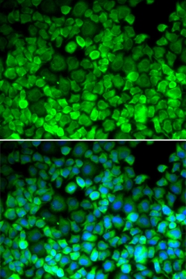
![TRF1 antibody [C3], C-term detects TRF1 protein at spindle by immunofluorescent analysis. Sample: U2OS cells were fixed in 4% paraformaldehyde at RT for 15 min. Green: TRF1 stained by TRF1 antibody [C3], C-term (GTX106349) diluted at 1:500. Blue: Fluoroshield with DAPI (GTX30920).](https://www.genetex.com/upload/website/prouct_img/normal/GTX106349/GTX106349_39701_20200212_ICC_IF_w_23060120_582.webp)
Product group Antibodies
TRF1 antibody [C3], C-termGTX106349
ApplicationsImmunoFluorescence, Western Blot, ImmunoCytoChemistry
ReactivityHuman
TargetTERF1
- SizePrice