

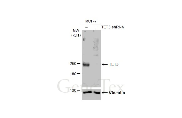
Non-transfected (–) and transfected (+) MCF-7 whole cell extracts (30 μg) were separated by 5% SDS-PAGE, and the membrane was blotted with TET3 antibody [HL2463] (GTX638781) diluted at 1:1000. The HRP-conjugated anti-rabbit IgG antibody (GTX213110-01) was used to detect the primary antibody, and the signal was developed with Trident ECL plus-Enhanced.
![Non-transfected (–) and transfected (+) 293T whole cell extracts (30 μg) were separated by 5% SDS-PAGE, and the membrane was blotted with TET3 antibody [HL2463] (GTX638781) diluted at 1:5000. The HRP-conjugated anti-rabbit IgG antibody (GTX213110-01) was used to detect the primary antibody.](https://www.genetex.com/upload/website/prouct_img/normal/GTX638781/GTX638781_T-45089_20240614_WB_multiple_B_24061802_726.webp "Non-transfected (–) and transfected (+) 293T whole cell extracts (30 μg) were separated by 5% SDS-PAGE, and the membrane was blotted with TET3 antibody [HL2463] (GTX638781) diluted at 1:5000. The HRP-conjugated anti-rabbit IgG antibody (GTX213110-01) was used to detect the primary antibody.")
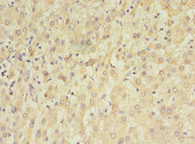
![TET3 antibody [HL2463] detects TET3 protein by immunohistochemical analysis. Sample: Paraffin-embedded human breast carcinoma. TET3 stained by TET3 antibody [HL2463] (GTX638781) diluted at 1:100. Antigen Retrieval: Citrate buffer, pH 6.0, 15 min](https://www.genetex.com/upload/website/prouct_img/normal/GTX638781/GTX638781_T-45089_20240626_IHC-P_24070822_112.webp "TET3 antibody [HL2463] detects TET3 protein by immunohistochemical analysis. Sample: Paraffin-embedded human breast carcinoma. TET3 stained by TET3 antibody [HL2463] (GTX638781) diluted at 1:100. Antigen Retrieval: Citrate buffer, pH 6.0, 15 min")
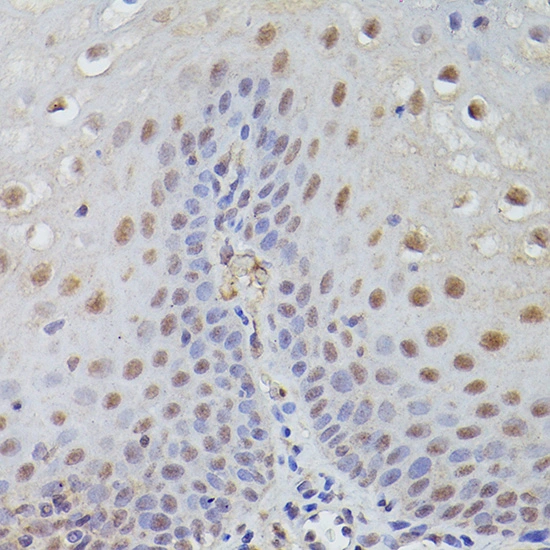
![TET3 antibody [HL2463] detects TET3 protein by immunohistochemical analysis. Sample: Paraffin-embedded mouse tissues. TET3 stained by TET3 antibody [HL2463] (GTX638781) diluted at 1:100. Antigen Retrieval: Citrate buffer, pH 6.0, 15 min](https://www.genetex.com/upload/website/prouct_img/normal/GTX638781/GTX638781_T-45089_20240626_IHC-P_multiple_M_24070822_739.webp "TET3 antibody [HL2463] detects TET3 protein by immunohistochemical analysis. Sample: Paraffin-embedded mouse tissues. TET3 stained by TET3 antibody [HL2463] (GTX638781) diluted at 1:100. Antigen Retrieval: Citrate buffer, pH 6.0, 15 min")
![TET3 antibody [HL2463] detects TET3 protein by immunohistochemical analysis. Sample: Paraffin-embedded rat tissues. TET3 stained by TET3 antibody [HL2463] (GTX638781) diluted at 1:100. Antigen Retrieval: Citrate buffer, pH 6.0, 15 min](https://www.genetex.com/upload/website/prouct_img/normal/GTX638781/GTX638781_T-45089_20240626_IHC-P_multiple_R_24070822_120.webp "TET3 antibody [HL2463] detects TET3 protein by immunohistochemical analysis. Sample: Paraffin-embedded rat tissues. TET3 stained by TET3 antibody [HL2463] (GTX638781) diluted at 1:100. Antigen Retrieval: Citrate buffer, pH 6.0, 15 min")
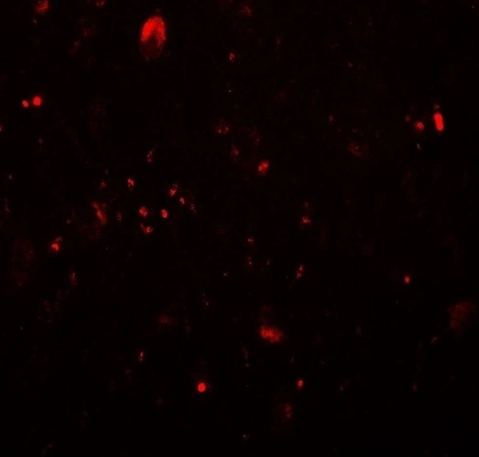
![Raw264.7 whole cell and nuclear extracts (30 μg) were separated by 5% SDS-PAGE, and the membrane was blotted with TET3 antibody [HL2463] (GTX638781) diluted at 1:1000. The HRP-conjugated anti-rabbit IgG antibody (GTX213110-01) was used to detect the primary antibody.](https://www.genetex.com/upload/website/prouct_img/normal/GTX638781/GTX638781_45460_20241220_WB_M_fraction_24122400_580.webp "Raw264.7 whole cell and nuclear extracts (30 μg) were separated by 5% SDS-PAGE, and the membrane was blotted with TET3 antibody [HL2463] (GTX638781) diluted at 1:1000. The HRP-conjugated anti-rabbit IgG antibody (GTX213110-01) was used to detect the primary antibody.")
Non-transfected (–) and transfected (+) MCF-7 whole cell extracts (30 μg) were separated by 5% SDS-PAGE, and the membrane was blotted with TET3 antibody [HL2463] (GTX638781) diluted at 1:1000. The HRP-conjugated anti-rabbit IgG antibody (GTX213110-01) was used to detect the primary antibody, and the signal was developed with Trident ECL plus-Enhanced.
TET3 antibody [HL2463]
GTX638781
ApplicationsWestern Blot, ImmunoHistoChemistry, ImmunoHistoChemistry Paraffin
Product group Antibodies
ReactivityHuman, Mouse, Rat
TargetTET3
Overview
- SupplierGeneTex
- Product NameTET3 antibody [HL2463]
- Delivery Days Customer7
- Application Supplier NoteWB: 1:500-1:3000. *Optimal dilutions/concentrations should be determined by the researcher.Not tested in other applications.
- ApplicationsWestern Blot, ImmunoHistoChemistry, ImmunoHistoChemistry Paraffin
- CertificationResearch Use Only
- ClonalityMonoclonal
- Clone IDHL2463
- Concentration1 mg/ml
- ConjugateUnconjugated
- Gene ID200424
- Target nameTET3
- Target descriptiontet methylcytosine dioxygenase 3
- Target synonymsBEFAHRS, hCG_40738, methylcytosine dioxygenase TET3, probable methylcytosine dioxygenase TET3, putative methylcytosine dioxygenase, ten-eleven translocation 3, tet oncogene family member 3
- HostRabbit
- IsotypeIgG
- Protein IDO43151
- Protein NameMethylcytosine dioxygenase TET3
- Scientific DescriptionMembers of the ten-eleven translocation (TET) gene family, including TET3, play a role in the DNA methylation process (Langemeijer et al., 2009 [PubMed 19923888]).[supplied by OMIM, Nov 2010]
- ReactivityHuman, Mouse, Rat
- Storage Instruction-20°C or -80°C,2°C to 8°C
- UNSPSC12352203
Datasheet
Related products
Product group Antibodies
Anti-TET3 Antibody144-64874
ApplicationsWestern Blot
ReactivityHuman, Mouse
TargetTET3
- SizePrice
Product group Antibodies

TET3 AntibodyLS-C748504
ApplicationsImmunoFluorescence
ReactivityHuman
TargetTET3
- SizePrice
![Non-transfected (–) and transfected (+) 293T whole cell extracts (30 μg) were separated by 5% SDS-PAGE, and the membrane was blotted with TET3 antibody [HL3009] (GTX640422) diluted at 1:5000. The HRP-conjugated anti-rabbit IgG antibody (GTX213110-01) was used to detect the primary antibody.](https://www.genetex.com/upload/website/prouct_img/normal/GTX640422/GTX640422_45509_20240823_WB_multiple_B_24090423_504.webp)
Product group Antibodies
TET3 antibody [HL3009]GTX640422
ApplicationsWestern Blot
ReactivityHuman
TargetTET3
- SizePrice
Product group Antibodies
TET3 AntibodyCSB-PA023398LA01HU
ApplicationsELISA, ImmunoHistoChemistry
ReactivityHuman
TargetTET3
- SizePrice

Product group Antibodies
Anti-TET3 AntibodyHPA050845
ApplicationsImmunoCytoChemistry, ImmunoHistoChemistry
ReactivityHuman
TargetTET3
- SizePrice
![Non-transfected (–) and transfected (+) 293T whole cell extracts (30 μg) were separated by 5% SDS-PAGE, and the membrane was blotted with TET3 antibody [N3C1], Internal (GTX121452) diluted at 1:5000. The HRP-conjugated anti-rabbit IgG antibody (GTX213110-01) was used to detect the primary antibody.](https://www.genetex.com/upload/website/prouct_img/normal/GTX121452/GTX121452_40464_20231229_WB_B_24010223_950.webp)
Product group Antibodies
References
TET3 antibody [N3C1], InternalGTX121452
ApplicationsWestern Blot
ReactivityHuman
TargetTET3
- SizePrice
![Non-transfected (–) and transfected (+) 293T whole cell extracts (30 μg) were separated by 5% SDS-PAGE, and the membrane was blotted with TET3 antibody [C3], C-term (GTX121453) diluted at 1:5000. The HRP-conjugated anti-rabbit IgG antibody (GTX213110-01) was used to detect the primary antibody, and the signal was developed with Trident ECL plus-Enhanced.](https://www.genetex.com/upload/website/prouct_img/normal/GTX121453/GTX121453_41570_20220617_WB_B_22062121_249.webp)
Product group Antibodies
References
TET3 antibody [C3], C-termGTX121453
ApplicationsImmunoFluorescence, ImmunoPrecipitation, Western Blot, ChIP Chromatin ImmunoPrecipitation, ImmunoCytoChemistry, ImmunoHistoChemistry
ReactivityBovine, Human, Mouse, Xenopus
TargetTET3
- SizePrice
Product group Antibodies
References
TET3 antibodyGTX00657
ApplicationsImmunoFluorescence, Western Blot, ImmunoCytoChemistry, ImmunoHistoChemistry, ImmunoHistoChemistry Paraffin
ReactivityHuman, Mouse
TargetTET3
- SizePrice
Product group Antibodies
TET3 antibodyGTX02579
ApplicationsWestern Blot, ELISA, ImmunoHistoChemistry, ImmunoHistoChemistry Paraffin
ReactivityHuman, Mouse, Rat
TargetTET3
- SizePrice