

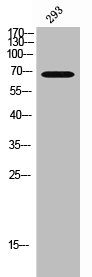
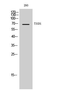
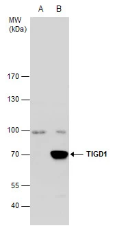
Western Blot analysis of 293 cells using TIGD1 Polyclonal Antibody
Western Blot analysis of 293 cells using TIGD1 Polyclonal Antibody
TIGD1 Antibody
CSB-PA008033
ApplicationsWestern Blot, ELISA
Product group Antibodies
ReactivityHuman
TargetTIGD1
Overview
- SupplierCusabio
- Product NameTIGD1 Antibody
- Delivery Days Customer20
- ApplicationsWestern Blot, ELISA
- CertificationResearch Use Only
- ClonalityPolyclonal
- ConjugateUnconjugated
- Gene ID200765
- Target nameTIGD1
- Target descriptiontigger transposable element derived 1
- Target synonymsEEYORE, tigger transposable element-derived protein 1
- HostRabbit
- IsotypeIgG
- Protein IDQ96MW7
- Protein NameTigger transposable element-derived protein 1
- ReactivityHuman
- Storage Instruction-20°C or -80°C
- UNSPSC41116161
Related products
Product group Antibodies
Anti-TIGD1 AntibodyA100740
ApplicationsWestern Blot, ELISA
ReactivityHuman
- SizePrice

Product group Antibodies
Anti-TIGD1 Antibody Picoband(r)A17411-1-CARRIER-FREE
ApplicationsFlow Cytometry, ImmunoFluorescence, Western Blot, ELISA, ImmunoCytoChemistry, ImmunoHistoChemistry
ReactivityHuman, Mouse, Rat
TargetTIGD1
- SizePrice
Product group Antibodies

Goat anti-TIGD1 / EEYOREEB06265
ApplicationsImmunoFluorescence, ELISA
ReactivityHuman
TargetTIGD1
- SizePrice

Product group Antibodies
Anti-TIGD1 AntibodyHPA041717
ApplicationsWestern Blot, ImmunoCytoChemistry, ImmunoHistoChemistry
ReactivityHuman
TargetTIGD1
- SizePrice
Product group Antibodies

ApplicationsWestern Blot, ELISA
ReactivityHuman
TargetTIGD1
- SizePrice
Product group Antibodies
TIGD1 antibody [C3], C-termGTX107727
ApplicationsWestern Blot
ReactivityHuman
TargetTIGD1
- SizePrice
Product group Antibodies
Anti-TIGD1 Antibody144-63998
ApplicationsWestern Blot
ReactivityHuman, Mouse, Rat
TargetTIGD1
- SizePrice