

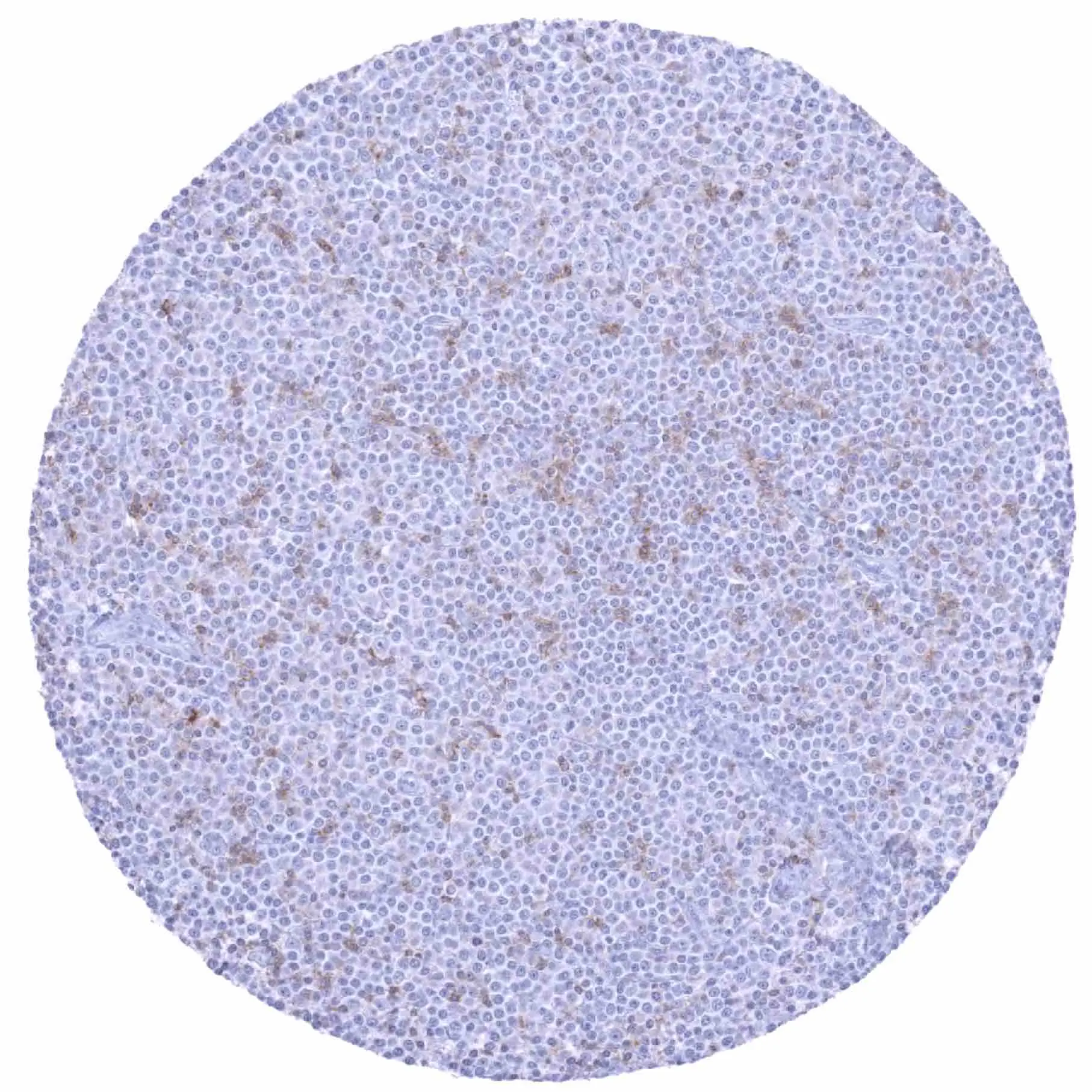
IHC-P analysis of human B-cell chronic lymphocytic leukemia (B-CLL) tissue using GTX639947 TIGIT antibody [HMV323] HistoMAX?. B-CLL with moderate to strong TIGIT positivity of a subset of tumor infiltrating lymphocytes.
![IHC-P analysis of human diffuse large B-cell lymphoma (DLBCL) from lymph node tissue using GTX639947 TIGIT antibody [HMV323] HistoMAX?. Diffuse large B-cell lymphoma with intense TIGIT positivity of numerous tumor infiltrating lymphocytes.](https://www.genetex.com/upload/website/prouct_img/normal/GTX639947/GTX639947_20240906_IHC-P_1_24090601_694.webp "IHC-P analysis of human diffuse large B-cell lymphoma (DLBCL) from lymph node tissue using GTX639947 TIGIT antibody [HMV323] HistoMAX?. Diffuse large B-cell lymphoma with intense TIGIT positivity of numerous tumor infiltrating lymphocytes.")
![IHC-P analysis of human follicular lymphoma (FL) tissue using GTX639947 TIGIT antibody [HMV323] HistoMAX?. Follicular B-cell lymphoma with strong TIGIT positivity of a portion of tumor infiltrating lymphocytes.](https://www.genetex.com/upload/website/prouct_img/normal/GTX639947/GTX639947_20240906_IHC-P_2_24090601_625.webp "IHC-P analysis of human follicular lymphoma (FL) tissue using GTX639947 TIGIT antibody [HMV323] HistoMAX?. Follicular B-cell lymphoma with strong TIGIT positivity of a portion of tumor infiltrating lymphocytes.")
![IHC-P analysis of human tonsil tissue using GTX639947 TIGIT antibody [HMV323] HistoMAX?. Numerous TIGIT positive lymphocytes. The strongest TIGIT staining appears in a subset of lymphocytes in the germinal centre.](https://www.genetex.com/upload/website/prouct_img/normal/GTX639947/GTX639947_20250214_IHC-P_25021323_718.webp "IHC-P analysis of human tonsil tissue using GTX639947 TIGIT antibody [HMV323] HistoMAX?. Numerous TIGIT positive lymphocytes. The strongest TIGIT staining appears in a subset of lymphocytes in the germinal centre.")
![IHC-P analysis of human Hodgkin lymphoma tissue using GTX639947 TIGIT antibody [HMV323] HistoMAX?. A distinct TIGIT positivity of a subset of non neoplastic lymphocytes surrounding tumor cells.](https://www.genetex.com/upload/website/prouct_img/normal/GTX639947/GTX639947_20250214_IHC-P_1_25021323_254.webp "IHC-P analysis of human Hodgkin lymphoma tissue using GTX639947 TIGIT antibody [HMV323] HistoMAX?. A distinct TIGIT positivity of a subset of non neoplastic lymphocytes surrounding tumor cells.")
IHC-P analysis of human B-cell chronic lymphocytic leukemia (B-CLL) tissue using GTX639947 TIGIT antibody [HMV323] HistoMAX?. B-CLL with moderate to strong TIGIT positivity of a subset of tumor infiltrating lymphocytes.
TIGIT antibody [HMV323] HistoMAX(tm)
GTX639947
ApplicationsImmunoHistoChemistry, ImmunoHistoChemistry Paraffin
Product group Antibodies
ReactivityHuman
TargetTIGIT
Overview
- SupplierGeneTex
- Product NameTIGIT antibody [HMV323] HistoMAX(tm)
- Delivery Days Customer7
- Application Supplier NoteIHC-P: 1:100-1:200. *Optimal dilutions/concentrations should be determined by the researcher.Not tested in other applications.
- ApplicationsImmunoHistoChemistry, ImmunoHistoChemistry Paraffin
- CertificationResearch Use Only
- ClonalityMonoclonal
- Clone IDHMV323
- Concentration0.25 mg/ml
- ConjugateUnconjugated
- Gene ID201633
- Target nameTIGIT
- Target descriptionT cell immunoreceptor with Ig and ITIM domains
- Target synonymsVSIG9, VSTM3, WUCAM, T-cell immunoreceptor with Ig and ITIM domains, V-set and immunoglobulin domain containing 9, V-set and immunoglobulin domain-containing protein 9, V-set and transmembrane domain containing 3, V-set and transmembrane domain-containing protein 3, VSIG9, VSTM3, Washington University cell adhesion molecule
- HostRabbit
- IsotypeIgG
- Protein IDQ495A1
- Protein NameT-cell immunoreceptor with Ig and ITIM domains
- Scientific DescriptionThis gene encodes a member of the PVR (poliovirus receptor) family of immunoglobin proteins. The product of this gene is expressed on several classes of T cells including follicular B helper T cells (TFH). The protein has been shown to bind PVR with high affinity; this binding is thought to assist interactions between TFH and dendritic cells to regulate T cell dependent B cell responses.[provided by RefSeq, Sep 2009]
- ReactivityHuman
- Storage Instruction-20°C or -80°C,2°C to 8°C
- UNSPSC41116161
Datasheet
Related products
Product group Antibodies
Anti-TIGIT AntibodyA306412
ApplicationsWestern Blot
ReactivityHuman, Mouse
- SizePrice
Product group Antibodies

Anti-TIGIT [4A5]Ab03137-1.1
ApplicationsFlow Cytometry, ELISA, Neutralisation/Blocking
ReactivityHuman
TargetTIGIT
- SizePrice
Product group Antibodies

ApplicationsELISA
ReactivityHuman
TargetTIGIT
- SizePrice
Product group Antibodies
TIGIT Polyclonal Antibody, AbBy Fluor-647 ConjugatedBS-43033R-BF647
ApplicationsWestern Blot
ReactivityHuman, Mouse, Rat
TargetTIGIT
- SizePrice
Product group Antibodies
TIGIT AntibodyCSB-PA675446LA01HU
ApplicationsImmunoFluorescence, ELISA
ReactivityHuman
TargetTIGIT
- SizePrice
Product group Antibodies
Tigit Polyclonal AntibodyCAC09330
ApplicationsImmunoFluorescence, ELISA
TargetTIGIT
- SizePrice
![Non-transfected (–) and transfected (+) 293T whole cell extracts (30 μg) were separated by 12% SDS-PAGE, and the membrane was blotted with TIGIT antibody [HL2655] (GTX639114) diluted at 1:30000. The HRP-conjugated anti-rabbit IgG antibody (GTX213110-01) was used to detect the primary antibody.](https://www.genetex.com/upload/website/prouct_img/normal/GTX639114/GTX639114_T-45194_20231110_WB_B_23111422_862.webp)
Product group Antibodies
TIGIT antibody [HL2655]GTX639114
ApplicationsWestern Blot
ReactivityHuman
TargetTIGIT
- SizePrice
![Various whole cell extracts (30 μg) were separated by 12% SDS-PAGE, and the membrane was blotted with TIGIT antibody [HL2657] (GTX639116) diluted at 1:1000. The HRP-conjugated anti-rabbit IgG antibody (GTX213110-01) was used to detect the primary antibody, and the signal was developed with Trident ECL plus-Enhanced. Corresponding RNA expression data for the same cell lines are based on Human Protein Atlas program.](https://www.genetex.com/upload/website/prouct_img/normal/GTX639116/GTX639116_T-45194_20231027_WB_TPM_watermark_23103019_943.webp)
Product group Antibodies
TIGIT antibody [HL2657]GTX639116
ApplicationsWestern Blot, ImmunoHistoChemistry, ImmunoHistoChemistry Paraffin
ReactivityHuman
TargetTIGIT
- SizePrice
![IHC-P analysis of human mucosa from appendix tissue using GTX639946 TIGIT antibody [HMV322] HistoMAX?. Distinct TIGIT staining of a subset of lymphocytes.](https://www.genetex.com/upload/website/prouct_img/normal/GTX639946/GTX639946_20240408_IHC-P_2_24040722_213.webp)
Product group Antibodies
TIGIT antibody [HMV322] HistoMAX(tm)GTX639946
ApplicationsImmunoHistoChemistry, ImmunoHistoChemistry Paraffin
ReactivityHuman
TargetTIGIT
- SizePrice