

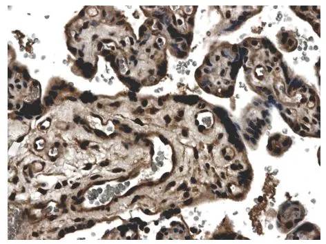
IHC-P analysis of formalin fixed human placenta tissue using GTX52488 TIMP3 antibody [7D3].
IHC-P analysis of formalin fixed human placenta tissue using GTX52488 TIMP3 antibody [7D3].
TIMP3 antibody [7D3]
GTX52488
ApplicationsWestern Blot, ImmunoHistoChemistry, ImmunoHistoChemistry Paraffin
Product group Antibodies
ReactivityHuman
TargetTIMP3
Overview
- SupplierGeneTex
- Product NameTIMP3 antibody [7D3]
- Delivery Days Customer9
- Application Supplier NoteWB: 1:100 - 1:1000. IHC-P: 1:50 - 1:200. *Optimal dilutions/concentrations should be determined by the researcher.Not tested in other applications.
- ApplicationsWestern Blot, ImmunoHistoChemistry, ImmunoHistoChemistry Paraffin
- CertificationResearch Use Only
- ClonalityMonoclonal
- Clone ID7D3
- Concentration200 ug/ml
- ConjugateUnconjugated
- Gene ID7078
- Target nameTIMP3
- Target descriptionTIMP metallopeptidase inhibitor 3
- Target synonymsHSMRK222, K222, K222TA2, SFD, metalloproteinase inhibitor 3, MIG-5 protein, TIMP-3, protein MIG-5, tissue inhibitor of metalloproteinases 3
- HostMouse
- IsotypeIgG1
- Protein IDP35625
- Protein NameMetalloproteinase inhibitor 3
- Scientific DescriptionThis gene belongs to the TIMP gene family. The proteins encoded by this gene family are inhibitors of the matrix metalloproteinases, a group of peptidases involved in degradation of the extracellular matrix (ECM). Expression of this gene is induced in response to mitogenic stimulation and this netrin domain-containing protein is localized to the ECM. Mutations in this gene have been associated with the autosomal dominant disorder Sorsbys fundus dystrophy. [provided by RefSeq, Jul 2008]
- ReactivityHuman
- Storage Instruction-20°C or -80°C,2°C to 8°C
- UNSPSC41116161
Datasheet
Related products

Product group Antibodies
Anti-TIMP3 AntibodyA97154
ApplicationsWestern Blot, ELISA
ReactivityHuman, Mouse, Rat
- SizePrice
Product group Antibodies
Anti-TIMP3 Antibody130-00010
ApplicationsWestern Blot, ELISA
ReactivityHuman
- SizePrice

Product group Antibodies
Anti-TIMP3 Antibody Picoband(r)A00477-CARRIER-FREE
ApplicationsWestern Blot, ELISA
ReactivityHuman, Mouse, Rat
TargetTIMP3
- SizePrice
Product group Antibodies
References
TIMP-3 Polyclonal AntibodyBS-0417R
ApplicationsImmunoFluorescence, Western Blot, ELISA, ImmunoCytoChemistry, ImmunoHistoChemistry, ImmunoHistoChemistry Frozen, ImmunoHistoChemistry Paraffin
ReactivityBovine, Equine, Human, Mouse, Porcine, Rabbit, Rat
TargetTIMP3
- SizePrice
Product group Antibodies
TIMP3 AntibodyCSB-PA004295
ApplicationsWestern Blot, ELISA
ReactivityHuman, Mouse, Rat
TargetTIMP3
- SizePrice
Product group Antibodies
TIMP3 Polyclonal AntibodyCAC14129
ApplicationsWestern Blot, ELISA
TargetTIMP3
- SizePrice
Product group Antibodies

TIMP3 Antibody (Preservative Free)LS-C149149
ApplicationsWestern Blot, ELISA
ReactivityHuman
TargetTIMP3
- SizePrice
![TIMP3 antibody [N1C3] detects TIMP3 protein by immunofluorescent analysis. Sample: DIV10 rat E18 primary cortical neuron cells were fixed in 4% paraformaldehyde at RT for 15 min. Green: TIMP3 stained by TIMP3 antibody [N1C3] (GTX100742) diluted at 1:500. Red: alpha Tubulin, stained by alpha Tubulin antibody [GT114] (GTX628802) diluted at 1:500. Blue: Fluoroshield with DAPI (GTX30920).](https://www.genetex.com/upload/website/prouct_img/normal/GTX100742/GTX100742_39721_20180503_ICC_IF_R_w_23060100_582.webp)
Product group Antibodies
TIMP3 antibody [N1C3]GTX100742
ApplicationsImmunoFluorescence, Western Blot, ImmunoCytoChemistry
ReactivityHuman, Mouse, Rat
TargetTIMP3
- SizePrice
Product group Antibodies
TIMP3 antibody, InternalGTX80547
ApplicationsFlow Cytometry, Western Blot, ImmunoHistoChemistry, ImmunoHistoChemistry Paraffin
ReactivityHuman, Mouse
TargetTIMP3
- SizePrice
![Immunofluorescent staining of human cell line BJ [Human fibroblast] shows localization to the Golgi apparatus & vesicles.](https://atlasantibodies.s3.amazonaws.com/images/icc/hpa068391-icc-1.jpg)
Product group Antibodies
Anti-TIMP3 AntibodyHPA068391
ApplicationsImmunoCytoChemistry
ReactivityHuman
TargetTIMP3
- SizePrice