

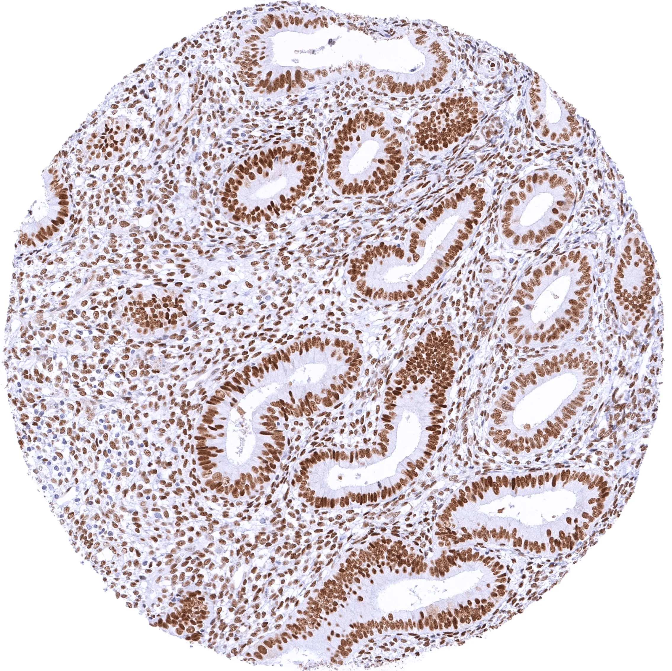
IHC-P analysis of human endometrium tissue using GTX04966 TLE1 antibody [MSVA-856R] HistoMAX?. Endometrium with moderate to strong TLE1 staining of epithelial and stromal cells.
![IHC-P analysis of human lymph node tissue using GTX04966 TLE1 antibody [MSVA-856R] HistoMAX?. Lymph node with weak to moderate TLE1 staining in a subset of lymphocytes predominantly in germinal centres.](https://www.genetex.com/upload/website/prouct_img/normal/GTX04966/GTX04966_20241028_IHC-P_1_24102820_420.webp "IHC-P analysis of human lymph node tissue using GTX04966 TLE1 antibody [MSVA-856R] HistoMAX?. Lymph node with weak to moderate TLE1 staining in a subset of lymphocytes predominantly in germinal centres.")
![IHC-P analysis of human synovial sarcoma tissue using GTX04966 TLE1 antibody [MSVA-856R] HistoMAX?. Synovial sarcoma with strong TLE1 positivity of all tumor cells.](https://www.genetex.com/upload/website/prouct_img/normal/GTX04966/GTX04966_20241028_IHC-P_24102820_568.webp "IHC-P analysis of human synovial sarcoma tissue using GTX04966 TLE1 antibody [MSVA-856R] HistoMAX?. Synovial sarcoma with strong TLE1 positivity of all tumor cells.")
IHC-P analysis of human endometrium tissue using GTX04966 TLE1 antibody [MSVA-856R] HistoMAX?. Endometrium with moderate to strong TLE1 staining of epithelial and stromal cells.
TLE1 antibody [MSVA-856R] HistoMAX(tm)
GTX04966
ApplicationsImmunoHistoChemistry, ImmunoHistoChemistry Paraffin
Product group Antibodies
ReactivityHuman
TargetTLE1
Overview
- SupplierGeneTex
- Product NameTLE1 antibody [MSVA-856R] HistoMAX(tm)
- Delivery Days Customer7
- Application Supplier NoteIHC-P: 1:100 - 1:200. *Optimal dilutions/concentrations should be determined by the researcher.Not tested in other applications.
- ApplicationsImmunoHistoChemistry, ImmunoHistoChemistry Paraffin
- CertificationResearch Use Only
- ClonalityMonoclonal
- Clone IDMSVA-856R
- Concentration66.66 ug/ml
- ConjugateUnconjugated
- Gene ID7088
- Target nameTLE1
- Target descriptionTLE family member 1, transcriptional corepressor
- Target synonymsESG, ESG1, GRG1, TLE-1, transducin-like enhancer protein 1, enhancer of split groucho-like protein 1, transducin like enhancer of split 1, transducin-like enhancer of split 1 (E(sp1) homolog, Drosophila)
- HostRabbit
- IsotypeIgG
- Protein IDQ04724
- Protein NameTransducin-like enhancer protein 1
- Scientific DescriptionEnables identical protein binding activity and transcription corepressor activity. Involved in negative regulation of I-kappaB kinase/NF-kappaB signaling; negative regulation of anoikis; and regulation of gene expression. Located in cytosol and nucleoplasm. Part of beta-catenin-TCF complex. [provided by Alliance of Genome Resources, Apr 2022]
- ReactivityHuman
- Storage Instruction-20°C or -80°C,2°C to 8°C
- UNSPSC41116161
Datasheet
Related products
Product group Antibodies
Anti-TLE1 AntibodyA35563
ApplicationsImmunoFluorescence, Western Blot, ImmunoHistoChemistry
ReactivityHuman, Mouse, Rat
- SizePrice
Product group Antibodies

TLE1 / TLE 1 AntibodyLS-C748412
ApplicationsWestern Blot
ReactivityHuman, Mouse
TargetTLE1
- SizePrice

Product group Antibodies
ApplicationsImmunoFluorescence, Western Blot, ImmunoCytoChemistry, ImmunoHistoChemistry
ReactivityHuman, Mouse, Rat
TargetTLE1
- SizePrice

Product group Antibodies
Anti-TLE1 AntibodyHPA071396
ApplicationsImmunoCytoChemistry
ReactivityHuman
TargetTLE1
- SizePrice
Product group Antibodies
TLE1 AntibodyCSB-PA717264ESR1HU
ApplicationsELISA
ReactivityHuman
TargetTLE1
- SizePrice
Product group Antibodies
TLE1 Recombinant Antibody, AbBy Fluor-488 ConjugatedBSM-61767R-BF488
ApplicationsImmunoFluorescence, Western Blot
ReactivityHuman, Mouse, Rat
TargetTLE1
- SizePrice
![TLE1 antibody [N2C1], Internal detects TLE1 protein by immunofluorescent analysis. Sample: DIV10 rat E18 primary cortical neuron cells were fixed in 4% paraformaldehyde at RT for 15 min. Green: TLE1 stained by TLE1 antibody [N2C1], Internal (GTX110092) diluted at 1:500. Red: beta Tubulin 3/ Tuj1, stained by beta Tubulin 3/ Tuj1 antibody [GT11710] (GTX631836) diluted at 1:500. Blue: Fluoroshield with DAPI (GTX30920).](https://www.genetex.com/upload/website/prouct_img/normal/GTX110092/GTX110092_40058_20181227_ICC_IF_R_w_23060500_841.webp)
Product group Antibodies
TLE1 antibody [N2C1], InternalGTX110092
ApplicationsImmunoFluorescence, Western Blot, ImmunoCytoChemistry
ReactivityHuman, Rat
TargetTLE1
- SizePrice
Product group Antibodies
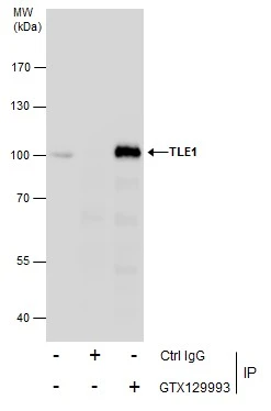
TLE1 antibodyGTX129993
ApplicationsImmunoPrecipitation, Western Blot, ImmunoHistoChemistry, ImmunoHistoChemistry Frozen
ReactivityHuman, Mouse
TargetTLE1
- SizePrice
Product group Antibodies
TLE1 antibody [ZM93]GTX01840
ApplicationsImmunoHistoChemistry, ImmunoHistoChemistry Paraffin
ReactivityHuman
TargetTLE1
- SizePrice