

TNF Receptor II antibody [HM102]
GTX27369
ApplicationsFlow Cytometry, ImmunoPrecipitation, Western Blot, ELISA, ImmunoHistoChemistry, ImmunoHistoChemistry Frozen, Other Application
Product group Antibodies
ReactivityMouse
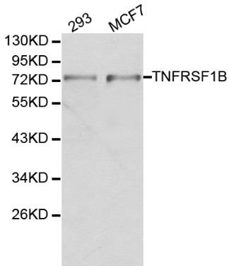
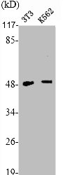
TargetTNFRSF1B
Overview
- SupplierGeneTex
- Product NameTNF Receptor II antibody [HM102]
- Delivery Days Customer9
- Application Supplier NoteFCM: 10microg/ml antibody in PBS/1% serum / 105 microglia cells. Activation: 2microg/ml. *Optimal dilutions/concentrations should be determined by the researcher.Not tested in other applications.
- ApplicationsFlow Cytometry, ImmunoPrecipitation, Western Blot, ELISA, ImmunoHistoChemistry, ImmunoHistoChemistry Frozen, Other Application
- CertificationResearch Use Only
- ClonalityMonoclonal
- Clone IDHM102
- Concentration100 ug/ml
- ConjugateUnconjugated
- Gene ID7133
- Target nameTNFRSF1B
- Target descriptionTNF receptor superfamily member 1B
- Target synonymsCD120b, TBPII, TNF-R-II, TNF-R75, TNFBR, TNFR1B, TNFR2, TNFR80, p75, p75TNFR, tumor necrosis factor receptor superfamily member 1B, TNF-R2, TNF-RII, p75 TNF receptor, p80 TNF-alpha receptor, tumor necrosis factor beta receptor, tumor necrosis factor binding protein 2, tumor necrosis factor receptor 2, tumor necrosis factor receptor type II
- HostRat
- IsotypeIgG2a
- Protein IDP20333
- Protein NameTumor necrosis factor receptor superfamily member 1B
- Scientific DescriptionThe protein encoded by this gene is a member of the TNF-receptor superfamily. This protein and TNF-receptor 1 form a heterocomplex that mediates the recruitment of two anti-apoptotic proteins, c-IAP1 and c-IAP2, which possess E3 ubiquitin ligase activity. The function of IAPs in TNF-receptor signalling is unknown, however, c-IAP1 is thought to potentiate TNF-induced apoptosis by the ubiquitination and degradation of TNF-receptor-associated factor 2, which mediates anti-apoptotic signals. Knockout studies in mice also suggest a role of this protein in protecting neurons from apoptosis by stimulating antioxidative pathways. [provided by RefSeq, Jul 2008]
- ReactivityMouse
- Storage Instruction2°C to 8°C
- UNSPSC41116161
Datasheet
Related products
Product group Antibodies
Anti-TNFRSF1B Antibody144-01095
ApplicationsWestern Blot
ReactivityHuman, Mouse, Rat
TargetTNFRSF1B
- SizePrice
Product group Antibodies
Anti-TNFRSF1B AntibodyA29315
ApplicationsWestern Blot, ImmunoHistoChemistry
ReactivityHuman
- SizePrice
Product group Antibodies

Anti-TNFR2 [5E6]AB03366-1.1-BT
ApplicationsELISA, Neutralisation/Blocking
ReactivityHuman
TargetTNFRSF1B
- SizePrice

Product group Antibodies
Anti-TNF Receptor II/TNFRSF1B Antibody Picoband(r)A01437-1-CARRIER-FREE
ApplicationsFlow Cytometry, Western Blot, ELISA
ReactivityHuman, Mouse, Rat
TargetTNFRSF1B
- SizePrice
Product group Antibodies
ApplicationsImmunoPrecipitation, Western Blot, ImmunoCytoChemistry, ImmunoHistoChemistry
TargetTNFRSF1B
- SizePrice
Product group Antibodies
TNFRSF1B AntibodyCSB-PA004309
ApplicationsImmunoFluorescence, Western Blot, ELISA, ImmunoHistoChemistry
ReactivityHuman, Mouse, Rat
TargetTNFRSF1B
- SizePrice
Product group Antibodies
TNF Receptor II antibody [22221]GTX10503
ApplicationsFlow Cytometry, ImmunoFluorescence, ImmunoCytoChemistry, Neutralisation/Blocking
ReactivityHuman
TargetTNFRSF1B
- SizePrice
Product group Antibodies
TNF Receptor II antibodyGTX31250
ApplicationsFunctional Assay, ImmunoFluorescence, Western Blot, ELISA, ImmunoCytoChemistry
ReactivityHuman
TargetTNFRSF1B
- SizePrice
Product group Antibodies
TNF Receptor II antibody [2/220]GTX42466
ApplicationsWestern Blot, ELISA, ImmunoHistoChemistry, ImmunoHistoChemistry Frozen, Neutralisation/Blocking
ReactivityHuman
TargetTNFRSF1B
- SizePrice