

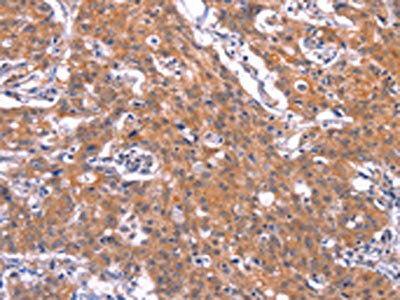
The image on the left is immunohistochemistry of paraffin-embedded Human gastic cancer tissue using CSB-PA057490(TNFRSF17 Antibody) at dilution 1/30, on the right is treated with synthetic peptide. (Original magnification: x200)
 at dilution 1/30, on the right is treated with synthetic peptide. (Original magnification: x200)")
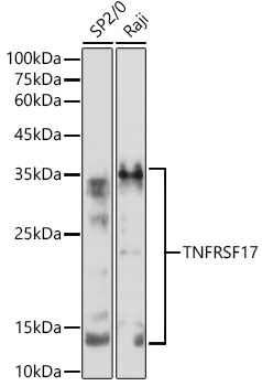
 at dilution 1/200, Secondary antibody: Goat anti rabbit IgG at 1/8000 dilution, Exposure time: 15 seconds")
The image on the left is immunohistochemistry of paraffin-embedded Human gastic cancer tissue using CSB-PA057490(TNFRSF17 Antibody) at dilution 1/30, on the right is treated with synthetic peptide. (Original magnification: x200)
TNFRSF17 Antibody
CSB-PA057490
ApplicationsWestern Blot, ELISA, ImmunoHistoChemistry
Product group Antibodies
ReactivityHuman, Mouse
TargetTNFRSF17
Overview
- SupplierCusabio
- Product NameTNFRSF17 Antibody
- Delivery Days Customer20
- ApplicationsWestern Blot, ELISA, ImmunoHistoChemistry
- CertificationResearch Use Only
- ClonalityPolyclonal
- ConjugateUnconjugated
- Gene ID608
- Target nameTNFRSF17
- Target descriptionTNF receptor superfamily member 17
- Target synonymsBCM, BCMA, CD269, TNFRSF13A, tumor necrosis factor receptor superfamily member 17, B cell maturation antigen, B-cell maturation factor, B-cell maturation protein
- HostRabbit
- IsotypeIgG
- Protein IDQ02223
- Protein NameTumor necrosis factor receptor superfamily member 17
- Scientific DescriptionThe protein encoded by this gene is a member of the TNF-receptor superfamily. This receptor is preferentially expressed in mature B lymphocytes, and may be important for B cell development and autoimmune response. This receptor has been shown to specifically bind to the tumor necrosis factor (ligand) superfamily, member 13b (TNFSF13B/TALL-1/BAFF), and to lead to NF-kappaB and MAPK8/JNK activation. This receptor also binds to various TRAF family members, and thus may transduce signals for cell survival and proliferation
- ReactivityHuman, Mouse
- Storage Instruction-20°C or -80°C
- UNSPSC41116161
Related products
Product group Antibodies
Anti-BCMA AntibodyA8765
ApplicationsImmunoFluorescence, Western Blot, ImmunoCytoChemistry
ReactivityHuman, Mouse, Rat
- SizePrice
Product group Antibodies

Anti-BCMA [B1]AB02727-23.159-BT
ApplicationsFlow Cytometry, ImmunoFluorescence, ELISA
ReactivityHuman
TargetTNFRSF17
- SizePrice
Product group Antibodies
Anti-TNFRSF17 (Center) Antibody102-22227
ApplicationsWestern Blot
TargetTNFRSF17
- SizePrice

Product group Antibodies
Anti-BCMA/TNFRSF17 Antibody Picoband(r)A01014-1-CARRIER-FREE
ApplicationsWestern Blot, ELISA, ImmunoHistoChemistry
ReactivityHuman
TargetTNFRSF17
- SizePrice
Product group Antibodies
BCMA Polyclonal AntibodyBS-3850R
ApplicationsFlow Cytometry, ImmunoFluorescence, Western Blot, ELISA, ImmunoCytoChemistry, ImmunoHistoChemistry, ImmunoHistoChemistry Frozen, ImmunoHistoChemistry Paraffin
ReactivityMouse, Rat
TargetTNFRSF17
- SizePrice
Product group Antibodies
TNFRSF17 Monoclonal AntibodyCAC13703
ApplicationsWestern Blot, ELISA, ImmunoHistoChemistry
TargetTNFRSF17
- SizePrice
Product group Antibodies

TNFRSF17 / BCMA AntibodyLS-C405921
ApplicationsWestern Blot, ELISA, ImmunoHistoChemistry
ReactivityHuman, Mouse
TargetTNFRSF17
- SizePrice
Product group Antibodies
BCMA antibody [Vicky-1]GTX17323
ApplicationsFlow Cytometry, ImmunoFluorescence, ImmunoCytoChemistry
ReactivityHuman
TargetTNFRSF17
- SizePrice