

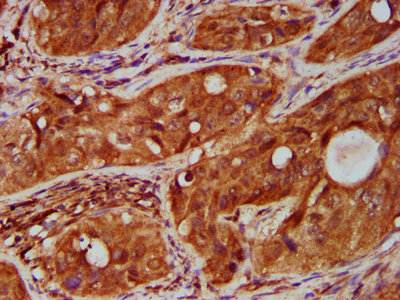
IHC image of CSB-PA023975LA01HU diluted at 1:500 and staining in paraffin-embedded human cervical cancer performed on a Leica BondTM system. After dewaxing and hydration, antigen retrieval was mediated by high pressure in a citrate buffer (pH 6.0). Section was blocked with 10% normal goat serum 30min at RT. Then primary antibody (1% BSA) was incubated at 4°C overnight. The primary is detected by a biotinylated secondary antibody and visualized using an HRP conjugated SP system.
. Section was blocked with 10% normal goat serum 30min at RT. Then primary antibody (1% BSA) was incubated at 4°C overnight. The primary is detected by a biotinylated secondary antibody and visualized using an HRP conjugated SP system.")
.")
IHC image of CSB-PA023975LA01HU diluted at 1:500 and staining in paraffin-embedded human cervical cancer performed on a Leica BondTM system. After dewaxing and hydration, antigen retrieval was mediated by high pressure in a citrate buffer (pH 6.0). Section was blocked with 10% normal goat serum 30min at RT. Then primary antibody (1% BSA) was incubated at 4°C overnight. The primary is detected by a biotinylated secondary antibody and visualized using an HRP conjugated SP system.
TNFRSF18 Antibody
CSB-PA023975LA01HU
ApplicationsImmunoFluorescence, ELISA, ImmunoHistoChemistry
Product group Antibodies
ReactivityHuman
TargetTNFRSF18
Overview
- SupplierCusabio
- Product NameTNFRSF18 Antibody
- Delivery Days Customer20
- ApplicationsImmunoFluorescence, ELISA, ImmunoHistoChemistry
- CertificationResearch Use Only
- ClonalityPolyclonal
- ConjugateUnconjugated
- Gene ID8784
- Target nameTNFRSF18
- Target descriptionTNF receptor superfamily member 18
- Target synonymsAITR, CD357, ENERGEN, GITR, GITR-D, tumor necrosis factor receptor superfamily member 18, TNF receptor superfamily activation-inducible protein, activation-inducible TNFR family receptor, glucocorticoid-induced TNFR-related protein
- HostRabbit
- IsotypeIgG
- Protein IDQ9Y5U5
- Protein NameTumor necrosis factor receptor superfamily member 18
- Scientific DescriptionReceptor for TNFSF18. Seems to be involved in interactions between activated T-lymphocytes and endothelial cells and in the regulation of T-cell receptor-mediated cell death. Mediated NF-kappa-B activation via the TRAF2/NIK pathway.
- ReactivityHuman
- Storage Instruction-20°C or -80°C
- UNSPSC41116161
Related products
Product group Antibodies
anti-GITR (human), pAbAG-25A-0017
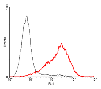
ApplicationsFlow Cytometry, ELISA
ReactivityHuman
TargetTNFRSF18
- SizePrice
Product group Antibodies

Anti-GITR [T10P2H9B9]Ab00727-1.1
ApplicationsWestern Blot, ELISA, ImmunoHistoChemistry
ReactivityHuman
TargetTNFRSF18
- SizePrice

Product group Antibodies
Anti-TNFRSF18 Antibody Picoband(r)A03125-1-CARRIER-FREE
ApplicationsWestern Blot
ReactivityHuman, Mouse, Rat
TargetTNFRSF18
- SizePrice

Product group Antibodies
Anti-TNFRSF18 AntibodyAMAB91487
ApplicationsWestern Blot
ReactivityHuman
TargetTNFRSF18
- SizePrice
Product group Antibodies

TNFRSF18 / GITR AntibodyLS-C681581
ApplicationsImmunoFluorescence, ELISA, ImmunoHistoChemistry, ImmunoHistoChemistry Paraffin
ReactivityHuman
TargetTNFRSF18
- SizePrice
Product group Antibodies
ApplicationsImmunoPrecipitation, Western Blot, ImmunoCytoChemistry, ImmunoHistoChemistry
ReactivityPorcine
TargetTNFRSF18
- SizePrice
![MJ whole cell and membrane extracts (30 μg) were separated by 12% SDS-PAGE, and the membrane was blotted with GITR antibody [HL3289] (GTX640943) diluted at 1:1000. The HRP-conjugated anti-rabbit IgG antibody (GTX213110-01) was used to detect the primary antibody.](https://www.genetex.com/upload/website/prouct_img/normal/GTX640943/GTX640943_T-45530_20240913_WB_Fraction_24091901_489.webp)
Product group Antibodies
GITR antibody [HL3289]GTX640943
ApplicationsWestern Blot
ReactivityHuman
TargetTNFRSF18
- SizePrice
Product group Antibodies
ApplicationsFlow Cytometry, ELISA
ReactivityHuman
- SizePrice