

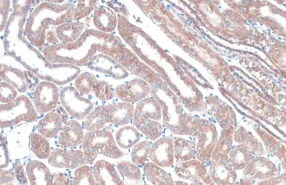
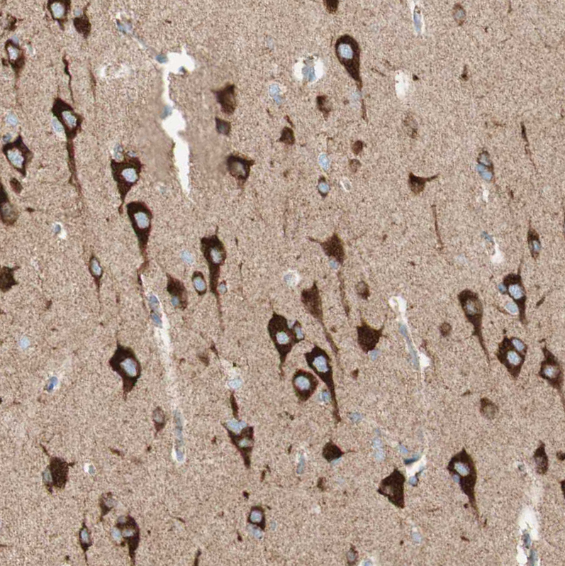
TOMM20 antibody detects TOMM20 protein at mitochondria by immunohistochemical analysis. Sample: Paraffin-embedded mouse kidney. TOMM20 stained by TOMM20 antibody (GTX133756) diluted at 1:615. Antigen Retrieval: Citrate buffer, pH 6.0, 15 min
![TOMM20 antibody detects TOMM20 protein at mitochondria by immunofluorescent analysis. Sample: HeLa cells were fixed in 4% paraformaldehyde at RT for 15 min. Green: TOMM20 stained by TOMM20 antibody (GTX133756) diluted at 1:500. Red: alpha Tubulin, a cytoskeleton marker, stained by alpha Tubulin antibody [GT114] (GTX628802) diluted at 1:1000. Blue: Fluoroshield with DAPI (GTX30920). Scale bar= 10μm.](https://www.genetex.com/upload/website/prouct_img/normal/GTX133756/GTX133756_44720_20220808_ICC_IF_22081423_208.webp "TOMM20 antibody detects TOMM20 protein at mitochondria by immunofluorescent analysis. Sample: HeLa cells were fixed in 4% paraformaldehyde at RT for 15 min. Green: TOMM20 stained by TOMM20 antibody (GTX133756) diluted at 1:500. Red: alpha Tubulin, a cytoskeleton marker, stained by alpha Tubulin antibody [GT114] (GTX628802) diluted at 1:1000. Blue: Fluoroshield with DAPI (GTX30920). Scale bar= 10μm.")
 diluted at 1:500. Antigen Retrieval: Citrate buffer, pH 6.0, 15 min")
 diluted at 1:500. Antigen Retrieval: Citrate buffer, pH 6.0, 15 min")
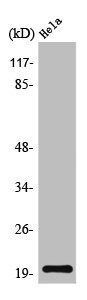
 was separated by 15% SDS-PAGE, and the membrane was blotted with TOMM20 antibody (GTX133756) diluted at 1:1000. The HRP-conjugated anti-rabbit IgG antibody (GTX213110-01) was used to detect the primary antibody.")
 were separated by 15% SDS-PAGE, and the membranes were blotted with TOMM20 antibody (GTX133756) diluted at 1:1000 and competitor's antibody (sc-11415) diluted at 1:1000. The HRP-conjugated anti-rabbit IgG antibody (GTX213110-01) was used to detect the primary antibody.")
 diluted at 1:500.
Antigen Retrieval: Citrate buffer, pH 6.0, 15 min")
 diluted at 1:500. Antigen Retrieval: Citrate buffer, pH 6.0, 15 min")
 was separated by 15% SDS-PAGE, and the membrane was blotted with TOMM20 antibody (GTX133756) diluted at 1:1000. The HRP-conjugated anti-rabbit IgG antibody (GTX213110-01) was used to detect the primary antibody.")
 diluted at 1:500.
Antigen Retrieval: Citrate buffer, pH 6.0, 15 min")
TOMM20 antibody detects TOMM20 protein at mitochondria by immunohistochemical analysis. Sample: Paraffin-embedded mouse kidney. TOMM20 stained by TOMM20 antibody (GTX133756) diluted at 1:615. Antigen Retrieval: Citrate buffer, pH 6.0, 15 min
TOMM20 antibody
GTX133756
ApplicationsImmunoFluorescence, Western Blot, ImmunoCytoChemistry, ImmunoHistoChemistry, ImmunoHistoChemistry Paraffin
Product group Antibodies
ReactivityHuman, Mouse, Rat
TargetTOMM20
Overview
- SupplierGeneTex
- Product NameTOMM20 antibody
- Delivery Days Customer9
- Application Supplier NoteWB: 1:500-1:3000. IHC-P: 1:100-1:1000. *Optimal dilutions/concentrations should be determined by the researcher.Not tested in other applications.
- ApplicationsImmunoFluorescence, Western Blot, ImmunoCytoChemistry, ImmunoHistoChemistry, ImmunoHistoChemistry Paraffin
- CertificationResearch Use Only
- ClonalityPolyclonal
- Concentration0.22 mg/ml
- ConjugateUnconjugated
- Gene ID9804
- Target nameTOMM20
- Target descriptiontranslocase of outer mitochondrial membrane 20
- Target synonymsMAS20, MOM19, TOM20, mitochondrial import receptor subunit TOM20 homolog, mitochondrial 20 kDa outer membrane protein, outer mitochondrial membrane receptor Tom20, translocase of outer mitochondrial membrane 20 homolog type II
- HostRabbit
- IsotypeIgG
- Protein IDQ15388
- Protein NameMitochondrial import receptor subunit TOM20 homolog
- Scientific DescriptionCentral component of the receptor complex responsible for the recognition and translocation of cytosolically synthesized mitochondrial preproteins. Together with TOM22 functions as the transit peptide receptor at the surface of the mitochondrion outer membrane and facilitates the movement of preproteins into the TOM40 translocation pore.
- ReactivityHuman, Mouse, Rat
- Storage Instruction-20°C or -80°C,2°C to 8°C
- UNSPSC41116161
Datasheet
Related products

Product group Antibodies
Anti-TOMM20 AntibodyA96710
ApplicationsWestern Blot, ELISA
ReactivityHuman, Mouse, Rat
- SizePrice

Product group Antibodies
Anti-TOMM20 Antibody Picoband(r)A04039-2-CARRIER-FREE
ApplicationsFlow Cytometry, ImmunoFluorescence, ImmunoPrecipitation, Western Blot, ELISA, ImmunoCytoChemistry, ImmunoHistoChemistry
ReactivityHuman, Mouse, Rat
TargetTOMM20
- SizePrice
Product group Antibodies
Anti-TOMM20 Antibody144-06774
ApplicationsImmunoFluorescence, Western Blot, ImmunoHistoChemistry
ReactivityHuman, Mouse, Rat
TargetTOMM20
- SizePrice
Product group Antibodies
TOMM20 Recombinant AntibodyBSM-61211R
ApplicationsFlow Cytometry, ImmunoFluorescence, Western Blot, ImmunoCytoChemistry, ImmunoHistoChemistry, ImmunoHistoChemistry Frozen, ImmunoHistoChemistry Paraffin
TargetTOMM20
- SizePrice
Product group Antibodies
TOMM20 AntibodyCSB-PA004315
ApplicationsWestern Blot, ELISA
ReactivityHuman, Mouse, Rat
TargetTOMM20
- SizePrice
Product group Antibodies
Tomm20 Polyclonal AntibodyCAC10576
ApplicationsImmunoFluorescence, Western Blot, ELISA, ImmunoHistoChemistry
ReactivityMouse
TargetTOMM20
- SizePrice
Product group Antibodies

TOMM20 Antibody (30-110 aa, Internal)LS-C386458
ApplicationsWestern Blot, ELISA
ReactivityHuman, Mouse, Rat
TargetTOMM20
- SizePrice
![IHC-P analysis of rat kidney tissue section using GTX00773 TOMM20 antibody [GT1136]. Dlution : 1:200](https://www.genetex.com/upload/website/prouct_img/normal/GTX00773/GTX00773_20191101_AP_003_2_w_23053121_479.webp)
Product group Antibodies
TOMM20 antibody [GT1136]GTX00773
ApplicationsImmunoFluorescence, ImmunoPrecipitation, Western Blot, ImmunoCytoChemistry, ImmunoHistoChemistry, ImmunoHistoChemistry Paraffin
ReactivityHuman, Mouse, Rat
TargetTOMM20
- SizePrice
Product group Antibodies
Anti-TOMM20 AntibodyHPA011562
ApplicationsWestern Blot, ImmunoCytoChemistry, ImmunoHistoChemistry
ReactivityHuman
TargetTOMM20
- SizePrice