

Topoisomerase I antibody [Mab1]
GTX12407
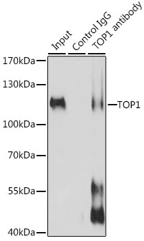
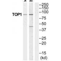
ApplicationsWestern Blot
Product group Antibodies
ReactivityHuman
TargetTOP1
Overview
- SupplierGeneTex
- Product NameTopoisomerase I antibody [Mab1]
- Delivery Days Customer9
- Application Supplier NoteWB: 1-2 microg/ml. *Optimal dilutions/concentrations should be determined by the researcher.Not tested in other applications.
- ApplicationsWestern Blot
- CertificationResearch Use Only
- ClonalityMonoclonal
- Clone IDMab1
- ConjugateUnconjugated
- Gene ID7150
- Target nameTOP1
- Target descriptionDNA topoisomerase I
- Target synonymsTOPI, DNA topoisomerase 1, Scl-70 antigen, topoisomerase (DNA) I, type I DNA topoisomerase
- HostMouse
- IsotypeIgG1
- Protein IDP11387
- Protein NameDNA topoisomerase 1
- Scientific DescriptionTopoisomerases are nuclear enzymes involved in a variety of cellular activities such as chromosome condensation, DNA replication, transcription, recombination and segregation at mitosis. Human topoisomerase I is a 100kD protein capable of relaxing positively and negatively supercoiled DNA by performing a transient single-stranded nick which is then re-ligated at the end of the reaction. It has been shown that the enzyme is located in regions of the genome that are undergoing active RNA synthesis, where it probably reduces superhelical stresses in the DNA, enabling RNA polymerase to function properly. In normal eukaryotic cells, DNA topoisomerase I does not show relevant fluctuations across the cell cycle, unlike DNA topoisomerase II alpha. Both DNA topoisomerases I and II have been found to be targets of autoantibodies in the sera of patients with certain autoimmune diseases such as systemic lupus erythematosus and also of some anti-tumor drugs and antibiotics. Elevated levels of DNA topoisomerase I, detected by 32P transfer assays, have been demonstrated in colorectal tumors compared with normal colon mucosa as a result of increased transcription or mRNA stability. Renal tumors were found not to show any differential levels compared with normal kidney. Topo I may be useful for the evaluation of DNA topoisomerase I expression in normal tissues, solid tumors and in further studies of ovarian, colorectal, cervical and prostatic tumors.
- ReactivityHuman
- Storage Instruction-20°C or -80°C,2°C to 8°C
- UNSPSC12352203
Datasheet
Related products
Product group Antibodies
Anti-TOP1 Antibody144-12524
ApplicationsImmunoPrecipitation, Western Blot, ImmunoHistoChemistry
ReactivityHuman, Mouse
TargetTOP1
- SizePrice
Product group Antibodies
Topoisomerase I antibodyGTX64445
ApplicationsImmunoPrecipitation, Western Blot, ImmunoHistoChemistry, ImmunoHistoChemistry Paraffin
ReactivityHuman, Mouse
TargetTOP1
- SizePrice
Product group Antibodies

Anti-DNA topoisomerase 1 [5C2]AB03245-1.1-BT
ApplicationsELISA
ReactivityHuman
TargetTOP1
- SizePrice
Product group Antibodies
Topoisomerase I antibody [23B11]GTX38947
ApplicationsWestern Blot
ReactivityHuman
TargetTOP1
- SizePrice
Product group Antibodies
ReactivityHuman
TargetTOP1
- SizePrice
Product group Antibodies
References
Topoisomerase I antibodyGTX129960
ApplicationsWestern Blot
ReactivityHuman
TargetTOP1
- SizePrice
Product group Antibodies
Topoisomerase I antibodyGTX130177
ApplicationsWestern Blot
ReactivityHuman, Rat
TargetTOP1
- SizePrice
Product group Antibodies
Topoisomerase I antibodyGTX130178
ApplicationsWestern Blot
ReactivityHuman
TargetTOP1
- SizePrice
Product group Antibodies
ApplicationsImmunoPrecipitation, Western Blot, ImmunoCytoChemistry, ImmunoHistoChemistry
TargetTOP1
- SizePrice
Product group Antibodies
Anti-TOP1 AntibodyA43790
ApplicationsWestern Blot
ReactivityHuman, Mouse, Rat
- SizePrice