

TOX antibody [N1C1] detects TOX protein at nucleus by immunohistochemical analysis. Sample: Paraffin-embedded mouse lymph node. TOX stained by TOX antibody [N1C1] (GTX115287) diluted at 1:500. Antigen Retrieval: Citrate buffer, pH 6.0, 15 min
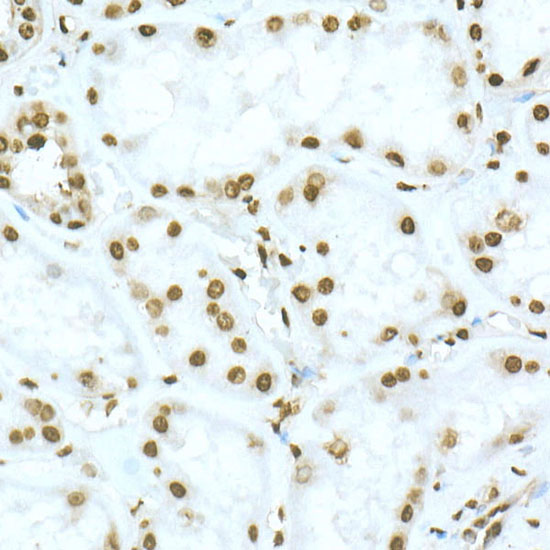
![TOX antibody [N1C1] detects TOX protein at nucleus by immunohistochemical analysis. Sample: Paraffin-embedded mouse spleen. TOX stained by TOX antibody [N1C1] (GTX115287) diluted at 1:500. Antigen Retrieval: Citrate buffer, pH 6.0, 15 min](https://www.genetex.com/upload/website/prouct_img/normal/GTX115287/GTX115287_44321_20210723_IHC-P_M_w_23060519_962.webp "TOX antibody [N1C1] detects TOX protein at nucleus by immunohistochemical analysis. Sample: Paraffin-embedded mouse spleen. TOX stained by TOX antibody [N1C1] (GTX115287) diluted at 1:500. Antigen Retrieval: Citrate buffer, pH 6.0, 15 min")
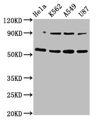
![Various tissue extracts (50 μg) were separated by 10% SDS-PAGE, and the membrane was blotted with TOX antibody [N1C1] (GTX115287) diluted at 1:1000. The HRP-conjugated anti-rabbit IgG antibody (GTX213110-01) was used to detect the primary antibody.](https://www.genetex.com/upload/website/prouct_img/normal/GTX115287/GTX115287_44272_20210528_WB_M_R_23100118_389.webp "Various tissue extracts (50 μg) were separated by 10% SDS-PAGE, and the membrane was blotted with TOX antibody [N1C1] (GTX115287) diluted at 1:1000. The HRP-conjugated anti-rabbit IgG antibody (GTX213110-01) was used to detect the primary antibody.")
![Various whole cell extracts (30 μg) were separated by 10% SDS-PAGE, and the membrane was blotted with TOX antibody [N1C1] (GTX115287) diluted at 1:1000. The HRP-conjugated anti-rabbit IgG antibody (GTX213110-01) was used to detect the primary antibody.](https://www.genetex.com/upload/website/prouct_img/normal/GTX115287/GTX115287_44272_20210409_WB_23100118_135.webp "Various whole cell extracts (30 μg) were separated by 10% SDS-PAGE, and the membrane was blotted with TOX antibody [N1C1] (GTX115287) diluted at 1:1000. The HRP-conjugated anti-rabbit IgG antibody (GTX213110-01) was used to detect the primary antibody.")
![Various whole cell extracts (30 μg) were separated by 10% SDS-PAGE, and the membrane was blotted with TOX antibody [N1C1] (GTX115287) diluted at 1:1000. The HRP-conjugated anti-rabbit IgG antibody (GTX213110-01) was used to detect the primary antibody. Corresponding RNA expression data for the same cell lines are based on Human Protein Atlas program.](https://www.genetex.com/upload/website/prouct_img/normal/GTX115287/GTX115287_43642_20231229_WB_TPM_watermark_24010223_552.webp "Various whole cell extracts (30 μg) were separated by 10% SDS-PAGE, and the membrane was blotted with TOX antibody [N1C1] (GTX115287) diluted at 1:1000. The HRP-conjugated anti-rabbit IgG antibody (GTX213110-01) was used to detect the primary antibody. Corresponding RNA expression data for the same cell lines are based on Human Protein Atlas program.")
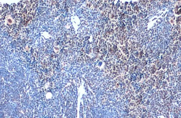
TOX antibody [N1C1] detects TOX protein at nucleus by immunohistochemical analysis. Sample: Paraffin-embedded mouse lymph node. TOX stained by TOX antibody [N1C1] (GTX115287) diluted at 1:500. Antigen Retrieval: Citrate buffer, pH 6.0, 15 min
TOX antibody [N1C1]
GTX115287
ApplicationsWestern Blot, ImmunoHistoChemistry, ImmunoHistoChemistry Paraffin
Product group Antibodies
ReactivityHuman, Mouse, Rat
TargetTOX
Overview
- SupplierGeneTex
- Product NameTOX antibody [N1C1]
- Delivery Days Customer9
- Application Supplier NoteWB: 1:500-1:3000. IHC-P: 1:100-1:1000. *Optimal dilutions/concentrations should be determined by the researcher.Not tested in other applications.
- ApplicationsWestern Blot, ImmunoHistoChemistry, ImmunoHistoChemistry Paraffin
- CertificationResearch Use Only
- ClonalityPolyclonal
- Concentration0.4 mg/ml
- ConjugateUnconjugated
- Gene ID9760
- Target nameTOX
- Target descriptionthymocyte selection associated high mobility group box
- Target synonymsTOX1, thymocyte selection-associated high mobility group box protein TOX, thymus high mobility group box protein TOX
- HostRabbit
- IsotypeIgG
- Protein IDO94900
- Protein NameThymocyte selection-associated high mobility group box protein TOX
- Scientific DescriptionThe protein encoded by this gene contains a HMG box DNA binding domain. HMG boxes are found in many eukaryotic proteins involved in chromatin assembly, transcription and replication. This protein may function to regulate T-cell development.
- ReactivityHuman, Mouse, Rat
- Storage Instruction-20°C or -80°C,2°C to 8°C
- UNSPSC41116161
Datasheet
Related products

Product group Antibodies
Anti-TOX Antibody Picoband(r)A08441-2-CARRIER-FREE
ApplicationsFlow Cytometry, Western Blot, ELISA, ImmunoHistoChemistry
ReactivityHuman, Mouse
TargetTOX
- SizePrice
Product group Antibodies
Anti-TOX AntibodyA10052
ApplicationsImmunoFluorescence, Western Blot, ImmunoCytoChemistry, ImmunoHistoChemistry
ReactivityHuman, Rat
- SizePrice
Product group Antibodies
Tox Polyclonal AntibodyCAC11318
ApplicationsWestern Blot, ELISA, ImmunoHistoChemistry
TargetTOX
- SizePrice
Product group Antibodies
TOX AntibodyCSB-PA024073LA01HU
ApplicationsWestern Blot, ELISA, ImmunoHistoChemistry
ReactivityHuman
TargetTOX
- SizePrice
Product group Antibodies

TOX AntibodyLS-C346202
ApplicationsImmunoFluorescence, Western Blot, ImmunoHistoChemistry
ReactivityHuman
TargetTOX
- SizePrice

Product group Antibodies
Anti-TOX AntibodyHPA073241
ApplicationsImmunoCytoChemistry
ReactivityHuman
TargetTOX
- SizePrice
![Various whole cell extracts (30 μg) were separated by 10% SDS-PAGE, and the membrane was blotted with TOX antibody [HL1809] (GTX637534) diluted at 1:1000. The HRP-conjugated anti-rabbit IgG antibody (GTX213110-01) was used to detect the primary antibody. Corresponding RNA expression data for the same cell lines are based on Human Protein Atlas program.](https://www.genetex.com/upload/website/prouct_img/normal/GTX637534/GTX637534_45313_20240223_WB_TPM_watermark_24030600_168.webp)
Product group Antibodies
TOX antibody [HL1809]GTX637534
ApplicationsWestern Blot
ReactivityHuman
TargetTOX
- SizePrice