

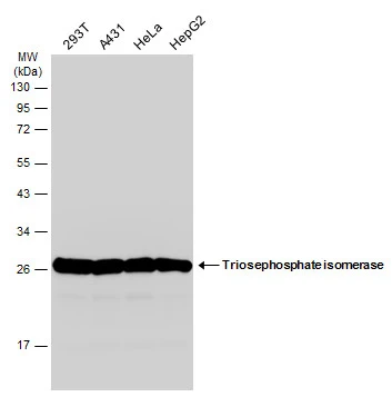
Various whole cell extracts (30 μg) were separated by 12% SDS-PAGE, and the membrane was blotted with Triosephosphate isomerase antibody [C2C3], C-term (GTX104618) diluted at 1:1000. The HRP-conjugated anti-rabbit IgG antibody (GTX213110-01) was used to detect the primary antibody.
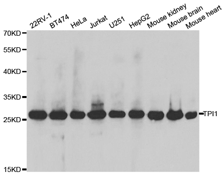
 were separated by 15% SDS-PAGE, and the membrane was blotted with Triosephosphate isomerase antibody (GTX104618) diluted by 1:1000. The HRP-conjugated anti-rabbit IgG antibody (GTX213110-01) was used to detect the primary antibody.")
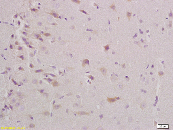
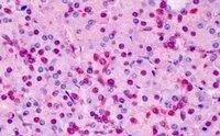
 antibody at 1:100 dilution.
Antigen Retrieval: Trilogy? (EDTA based, pH 8.0) buffer, 15min")
 were separated by 15% SDS-PAGE, and the membrane was blotted with Triosephosphate isomerase antibody (GTX104618) diluted by 1:1000. The HRP-conjugated anti-rabbit IgG antibody (GTX213110-01) was used to detect the primary antibody.")
![Triosephosphate isomerase antibody [C2C3], C-term detects Triosephosphate isomerase protein at cytoplasm by immunofluorescent analysis. Sample: HeLa cells were fixed in 4% paraformaldehyde at RT for 15 min. Green: Triosephosphate isomerase stained by Triosephosphate isomerase antibody [C2C3], C-term (GTX104618) diluted at 1:500. Blue: Hoechst 33342 staining. Scale bar= 10μm.](https://www.genetex.com/upload/website/prouct_img/normal/GTX104618/GTX104618_43117_20180307_ICC_IF_w_23060120_945.webp "Triosephosphate isomerase antibody [C2C3], C-term detects Triosephosphate isomerase protein at cytoplasm by immunofluorescent analysis. Sample: HeLa cells were fixed in 4% paraformaldehyde at RT for 15 min. Green: Triosephosphate isomerase stained by Triosephosphate isomerase antibody [C2C3], C-term (GTX104618) diluted at 1:500. Blue: Hoechst 33342 staining. Scale bar= 10μm.")
![Non-transfected (–) and transfected (+) 293T whole cell extracts (30 μg) were separated by 12% SDS-PAGE, and the membrane was blotted with Triosephosphate isomerase antibody [C2C3], C-term (GTX104618) diluted at 1:3000. The HRP-conjugated anti-rabbit IgG antibody (GTX213110-01) was used to detect the primary antibody.](https://www.genetex.com/upload/website/prouct_img/normal/GTX104618/GTX104618_41752_20161020_WB_shRNA_watermark_w_23060120_117.webp "Non-transfected (–) and transfected (+) 293T whole cell extracts (30 μg) were separated by 12% SDS-PAGE, and the membrane was blotted with Triosephosphate isomerase antibody [C2C3], C-term (GTX104618) diluted at 1:3000. The HRP-conjugated anti-rabbit IgG antibody (GTX213110-01) was used to detect the primary antibody.")

Various whole cell extracts (30 μg) were separated by 12% SDS-PAGE, and the membrane was blotted with Triosephosphate isomerase antibody [C2C3], C-term (GTX104618) diluted at 1:1000. The HRP-conjugated anti-rabbit IgG antibody (GTX213110-01) was used to detect the primary antibody.
Triosephosphate isomerase antibody [C2C3], C-term
GTX104618
ApplicationsImmunoFluorescence, Western Blot, ELISA, ImmunoCytoChemistry, ImmunoHistoChemistry, ImmunoHistoChemistry Paraffin
Product group Antibodies
ReactivityHuman, Mouse, Rat
TargetTPI1
Overview
- SupplierGeneTex
- Product NameTriosephosphate isomerase antibody [C2C3], C-term
- Delivery Days Customer9
- Application Supplier NoteWB: 1:500-1:3000. ICC/IF: 1:100-1:1000. IHC-P: 1:100-1:1000. *Optimal dilutions/concentrations should be determined by the researcher.Not tested in other applications.
- ApplicationsImmunoFluorescence, Western Blot, ELISA, ImmunoCytoChemistry, ImmunoHistoChemistry, ImmunoHistoChemistry Paraffin
- CertificationResearch Use Only
- ClonalityPolyclonal
- Concentration0.27 mg/ml
- ConjugateUnconjugated
- Gene ID7167
- Target nameTPI1
- Target descriptiontriosephosphate isomerase 1
- Target synonymsHEL-S-49, TIM, TPI, TPID, triosephosphate isomerase, epididymis secretory protein Li 49, methylglyoxal synthase, triose-phosphate isomerase
- HostRabbit
- IsotypeIgG
- Protein IDP60174
- Protein NameTriosephosphate isomerase
- Scientific DescriptionThis gene encodes an enzyme, consisting of two identical proteins, which catalyzes the isomerization of glyceraldehydes 3-phosphate (G3P) and dihydroxy-acetone phosphate (DHAP) in glycolysis and gluconeogenesis. Mutations in this gene are associated with triosephosphate isomerase deficiency. Pseudogenes have been identified on chromosomes 1, 4, 6 and 7. Alternative splicing results in multiple transcript variants. [provided by RefSeq]
- ReactivityHuman, Mouse, Rat
- Storage Instruction-20°C or -80°C,2°C to 8°C
- UNSPSC41116161
Datasheet
Related products
Product group Antibodies
Anti-TPI1 AntibodyA30927
ApplicationsImmunoFluorescence, Western Blot, ImmunoHistoChemistry
ReactivityHuman, Mouse, Rat
- SizePrice

Product group Antibodies
Anti-TPI1 Antibody Picoband(r)A02559-2-CARRIER-FREE
ApplicationsFlow Cytometry, Western Blot, ELISA
ReactivityHuman, Mouse, Rat
TargetTPI1
- SizePrice
Product group Antibodies
Anti-TPI1 Antibody144-02579
ApplicationsImmunoFluorescence, Western Blot, ImmunoHistoChemistry
ReactivityHuman, Mouse
TargetTPI1
- SizePrice
Product group Antibodies

Triosephosphate isomerase AntibodyABX431681
ApplicationsFlow Cytometry, Western Blot, ELISA, ImmunoHistoChemistry
- SizePrice
Product group Antibodies
ApplicationsImmunoFluorescence, Western Blot, ELISA, ImmunoCytoChemistry, ImmunoHistoChemistry, ImmunoHistoChemistry Frozen, ImmunoHistoChemistry Paraffin
ReactivityBovine, Canine, Chicken, Equine, Human, Mouse, Porcine, Rabbit, Rat, Sheep
TargetTPI1
- SizePrice
Product group Antibodies
TPI1 AntibodyCSB-PA13499A0RB
ApplicationsWestern Blot, ELISA, ImmunoHistoChemistry
ReactivityHuman, Mouse
TargetTPI1
- SizePrice
Product group Antibodies
References

ApplicationsWestern Blot, ELISA, ImmunoHistoChemistry
ReactivityBovine, Canine, Human, Mouse, Rat
TargetTPI1
- SizePrice
Product group Antibodies
TPI1 Polyclonal AntibodyCAC14655
ApplicationsWestern Blot, ELISA, ImmunoHistoChemistry
ReactivityMouse
TargetTPI1
- SizePrice
Product group Antibodies

TPI1 / TPI AntibodyLS-C482377
ApplicationsImmunoFluorescence, Western Blot, ImmunoCytoChemistry
ReactivityHuman, Mouse, Rat
TargetTPI1
- SizePrice
Product group Antibodies
ApplicationsWestern Blot, ImmunoHistoChemistry, ImmunoHistoChemistry Paraffin
ReactivityHuman, Mouse
TargetTPI1
- SizePrice