

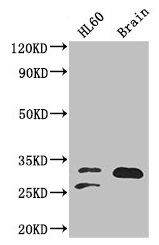
Western Blot Positive WB detected in: HL60 whole cell lysate, Mouse brain tissue All lanes: TPI1 antibody at 2microg/ml Secondary Goat polyclonal to rabbit IgG at 1/50000 dilution Predicted band size: 31, 27, 18 kDa Observed band size: 31, 27 kDa

Western Blot Positive WB detected in: HL60 whole cell lysate, Mouse brain tissue All lanes: TPI1 antibody at 2microg/ml Secondary Goat polyclonal to rabbit IgG at 1/50000 dilution Predicted band size: 31, 27, 18 kDa Observed band size: 31, 27 kDa
TPI1 Antibody
CSB-PA13499A0RB
ApplicationsWestern Blot, ELISA, ImmunoHistoChemistry
Product group Antibodies
ReactivityHuman, Mouse
TargetTPI1
Overview
- SupplierCusabio
- Product NameTPI1 Antibody
- Delivery Days Customer20
- ApplicationsWestern Blot, ELISA, ImmunoHistoChemistry
- CertificationResearch Use Only
- ClonalityPolyclonal
- ConjugateUnconjugated
- Gene ID7167
- Target nameTPI1
- Target descriptiontriosephosphate isomerase 1
- Target synonymsHEL-S-49, TIM, TPI, TPID, triosephosphate isomerase, epididymis secretory protein Li 49, methylglyoxal synthase, triose-phosphate isomerase
- HostRabbit
- IsotypeIgG
- Protein IDP60174
- Protein NameTriosephosphate isomerase
- Scientific Descriptioncytosol, extracellular exosome, extracellular space, nucleus, triose-phosphate isomerase activity, ubiquitin protein ligase binding, canonical glycolysis, gluconeogenesis, glyceraldehyde-3-phosphate biosynthetic process, glycerol catabolic process
- ReactivityHuman, Mouse
- Storage Instruction-20°C or -80°C
- UNSPSC41116161
Related products
Product group Antibodies
Anti-TPI1 AntibodyA30927
ApplicationsImmunoFluorescence, Western Blot, ImmunoHistoChemistry
ReactivityHuman, Mouse, Rat
- SizePrice

Product group Antibodies
Anti-TPI1 Antibody Picoband(r)A02559-2-CARRIER-FREE
ApplicationsFlow Cytometry, Western Blot, ELISA
ReactivityHuman, Mouse, Rat
TargetTPI1
- SizePrice
Product group Antibodies
Anti-TPI1 Antibody144-02579
ApplicationsImmunoFluorescence, Western Blot, ImmunoHistoChemistry
ReactivityHuman, Mouse
TargetTPI1
- SizePrice
Product group Antibodies

Triosephosphate isomerase AntibodyABX431681
ApplicationsFlow Cytometry, Western Blot, ELISA, ImmunoHistoChemistry
- SizePrice
Product group Antibodies
ApplicationsImmunoFluorescence, Western Blot, ELISA, ImmunoCytoChemistry, ImmunoHistoChemistry, ImmunoHistoChemistry Frozen, ImmunoHistoChemistry Paraffin
ReactivityBovine, Canine, Chicken, Equine, Human, Mouse, Porcine, Rabbit, Rat, Sheep
TargetTPI1
- SizePrice
Product group Antibodies
References

ApplicationsWestern Blot, ELISA, ImmunoHistoChemistry
ReactivityBovine, Canine, Human, Mouse, Rat
TargetTPI1
- SizePrice
Product group Antibodies
TPI1 Polyclonal AntibodyCAC14655
ApplicationsWestern Blot, ELISA, ImmunoHistoChemistry
ReactivityMouse
TargetTPI1
- SizePrice
Product group Antibodies

TPI1 / TPI AntibodyLS-C482377
ApplicationsImmunoFluorescence, Western Blot, ImmunoCytoChemistry
ReactivityHuman, Mouse, Rat
TargetTPI1
- SizePrice
Product group Antibodies
Anti-TPI1 AntibodyHPA050924
ApplicationsWestern Blot, ImmunoCytoChemistry, ImmunoHistoChemistry
ReactivityHuman
TargetTPI1
- SizePrice
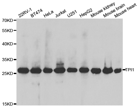
![Various whole cell extracts (30 μg) were separated by 12% SDS-PAGE, and the membrane was blotted with Triosephosphate isomerase antibody [C2C3], C-term (GTX104618) diluted at 1:1000. The HRP-conjugated anti-rabbit IgG antibody (GTX213110-01) was used to detect the primary antibody.](https://www.genetex.com/upload/website/prouct_img/normal/GTX104618/GTX104618_43117_20180216_WB_w_23060120_185.webp)
Product group Antibodies
ApplicationsImmunoFluorescence, Western Blot, ELISA, ImmunoCytoChemistry, ImmunoHistoChemistry, ImmunoHistoChemistry Paraffin
ReactivityHuman, Mouse, Rat
TargetTPI1
- SizePrice