

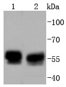
Lane 1: 293T Cells; Lane 2: Hela Cells; Probed with TRK fused gene (5C1) Monoclonal Antibody (bsm-52676R) at 1:1000 overnight at 4°C followed by a conjugated secondary antibody for 60 minutes at 37°C.
 staining with TRK fused gene (5C1) Monoclonal Antibody (bsm-52676R) at 1:100 in HeLa cells (green). The nuclear counterstain is DAPI (blue). Cells were fixed in paraformaldehyde, permeabilized with 0.25% Triton X100/PBS.")
 staining with TRK fused gene (5C1) Monoclonal Antibody (bsm-52676R) at 1:100 in A549 cells (green). The nuclear counterstain is DAPI (blue). Cells were fixed in paraformaldehyde, permeabilized with 0.25% Triton X100/PBS.")
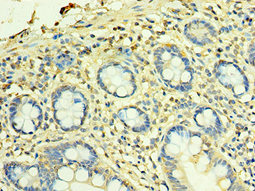
 Monoclonal Antibody (bsm-52676R) at 1:100, overnight at 4°C, followed by a conjugated secondary antibody and DAB staining. Counterstained with hematoxylin.")
 Monoclonal Antibody (bsm-52676R) at 1:100, overnight at 4°C, followed by a conjugated secondary antibody and DAB staining. Counterstained with hematoxylin.")
 Monoclonal Antibody (bsm-52676R) at 1:100, overnight at 4°C, followed by a conjugated secondary antibody and DAB staining. Counterstained with hematoxylin.")
 Monoclonal Antibody (bsm-52676R) at 1:100, overnight at 4°C, followed by a conjugated secondary antibody and DAB staining. Counterstained with hematoxylin.")
Lane 1: 293T Cells; Lane 2: Hela Cells; Probed with TRK fused gene (5C1) Monoclonal Antibody (bsm-52676R) at 1:1000 overnight at 4°C followed by a conjugated secondary antibody for 60 minutes at 37°C.
TFG Recombinant Antibody
BSM-52676R
ApplicationsImmunoFluorescence, Western Blot, ImmunoCytoChemistry, ImmunoHistoChemistry, ImmunoHistoChemistry Frozen, ImmunoHistoChemistry Paraffin
Product group Antibodies
ReactivityHuman, Mouse, Rat
TargetTFG
Overview
- SupplierBioss
- Product NameTFG Recombinant Antibody
- Delivery Days Customer16
- ApplicationsImmunoFluorescence, Western Blot, ImmunoCytoChemistry, ImmunoHistoChemistry, ImmunoHistoChemistry Frozen, ImmunoHistoChemistry Paraffin
- Applications SupplierWB(1:300-5000), IHC-P(1:200-400), IHC-F(1:100-500), IF(), ICC/IF(1:50-100)
- CertificationResearch Use Only
- ClonalityMonoclonal
- Concentration1 ug/ul
- ConjugateUnconjugated
- Gene ID10342
- Target nameTFG
- Target descriptiontrafficking from ER to golgi regulator
- Target synonymsHMSNP, SPG57, TF6, TRKT3, protein TFG, TRK-fused, TRK-fused gene protein, TRKT3 oncogene
- HostRabbit
- IsotypeIgG
- Protein IDQ92734
- Protein NameProtein TFG
- ReactivityHuman, Mouse, Rat
- Storage Instruction-20°C
- UNSPSC41116161
Datasheet
Related products

Product group Antibodies
Anti-TRK fused gene/TFG Antibody Picoband(r)A02870-3-CARRIER-FREE
ApplicationsImmunoFluorescence, Western Blot, ELISA, ImmunoCytoChemistry, ImmunoHistoChemistry
ReactivityHuman, Mouse, Rat
TargetTFG
- SizePrice
Product group Antibodies
Anti-TFG Antibody144-07769
ApplicationsWestern Blot, ImmunoHistoChemistry
ReactivityHuman, Mouse, Rat
TargetTFG
- SizePrice
Product group Antibodies
TFG AntibodyCSB-PA852902ESR1HU
ApplicationsELISA, ImmunoHistoChemistry
ReactivityHuman
TargetTFG
- SizePrice
Product group Antibodies

TFG AntibodyLS-C409319
ApplicationsWestern Blot, ImmunoHistoChemistry
ReactivityHuman, Mouse, Rat
TargetTFG
- SizePrice
![WB analysis of Jurkat cell lysate using GTX00523 TFG antibody [TFG-03].](https://www.genetex.com/upload/website/prouct_img/normal/GTX00523/GTX00523_20191025_AP_001_61_w_23053121_839.webp)
Product group Antibodies
TFG antibody [TFG-03]GTX00523
ApplicationsImmunoFluorescence, ImmunoPrecipitation, Western Blot, ImmunoCytoChemistry
ReactivityHuman
TargetTFG
- SizePrice

Product group Antibodies
Anti-TFG AntibodyHPA052206
ApplicationsWestern Blot, ImmunoCytoChemistry
ReactivityHuman
TargetTFG
- SizePrice