

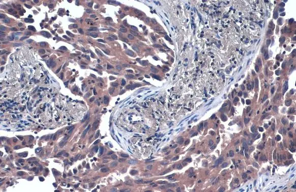
TSG101 antibody [4A10] detects TSG101 protein at cytoplasm by immunohistochemical analysis. Sample: Paraffin-embedded human lung cancer. TSG101 stained by TSG101 antibody [4A10] (GTX70255) diluted at 1:50. Antigen Retrieval: Citrate buffer, pH 6.0, 15 min
![Non-transfected (–) and transfected (+) 293T whole cell extracts (30 μg) were separated by 10% SDS-PAGE, and the membrane was blotted with TSG101 antibody [4A10] (GTX70255) diluted at 1:500. The HRP-conjugated anti-mouse IgG antibody (GTX213111-01) was used to detect the primary antibody.](https://www.genetex.com/upload/website/prouct_img/normal/GTX70255/GTX70255_41449_20161201_WB_shRNA_watermark_w_23061221_273.webp "Non-transfected (–) and transfected (+) 293T whole cell extracts (30 μg) were separated by 10% SDS-PAGE, and the membrane was blotted with TSG101 antibody [4A10] (GTX70255) diluted at 1:500. The HRP-conjugated anti-mouse IgG antibody (GTX213111-01) was used to detect the primary antibody.")
![TSG101 antibody [4A10] detects TSG101 protein at cytoplasm by immunohistochemical analysis. Sample: Paraffin-embedded human ovarian cancer. TSG101 stained by TSG101 antibody [4A10] (GTX70255) diluted at 1:100. Antigen Retrieval: Citrate buffer, pH 6.0, 15 min](https://www.genetex.com/upload/website/prouct_img/normal/GTX70255/GTX70255_43284_20181025_IHC-P_1_w_23061221_566.webp "TSG101 antibody [4A10] detects TSG101 protein at cytoplasm by immunohistochemical analysis. Sample: Paraffin-embedded human ovarian cancer. TSG101 stained by TSG101 antibody [4A10] (GTX70255) diluted at 1:100. Antigen Retrieval: Citrate buffer, pH 6.0, 15 min")

![Various tissue extracts (50 μg) were separated by 10% SDS-PAGE, and the membrane was blotted with TSG101 antibody [4A10] (GTX70255) diluted at 1:500. The HRP-conjugated anti-mouse IgG antibody (GTX213111-01) was used to detect the primary antibody.](https://www.genetex.com/upload/website/prouct_img/normal/GTX70255/GTX70255_43344_20190315_WB_M_tissue_w_23061221_184.webp "Various tissue extracts (50 μg) were separated by 10% SDS-PAGE, and the membrane was blotted with TSG101 antibody [4A10] (GTX70255) diluted at 1:500. The HRP-conjugated anti-mouse IgG antibody (GTX213111-01) was used to detect the primary antibody.")
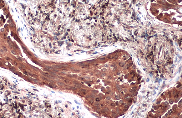
![TSG101 antibody [4A10] detects TSG101 protein at cytoplasm by immunohistochemical analysis. Sample: Paraffin-embedded human breast carcinoma. TSG101 stained by TSG101 antibody [4A10] (GTX70255) diluted at 1:100. Antigen Retrieval: Citrate buffer, pH 6.0, 15 min](https://www.genetex.com/upload/website/prouct_img/normal/GTX70255/GTX70255_43284_20181025_IHC-P_w_23061221_587.webp "TSG101 antibody [4A10] detects TSG101 protein at cytoplasm by immunohistochemical analysis. Sample: Paraffin-embedded human breast carcinoma. TSG101 stained by TSG101 antibody [4A10] (GTX70255) diluted at 1:100. Antigen Retrieval: Citrate buffer, pH 6.0, 15 min")
![TSG101 antibody [4A10] (GTX70255) detects TSG101 protein by flow cytometry analysis. Sample: THP-1 cell. Black: Unlabelled sample was used as a control. Red: TSG101 antibody [4A10] (GTX70255) dilution: 1:25. Acquisition of 20,000 events were collected using a Dylight 488-conjugated secondary antibody for FACS analysis.](https://www.genetex.com/upload/website/prouct_img/normal/GTX70255/GTX70255_43383_20190201_FACS_w_23061221_980.webp "TSG101 antibody [4A10] (GTX70255) detects TSG101 protein by flow cytometry analysis. Sample: THP-1 cell. Black: Unlabelled sample was used as a control. Red: TSG101 antibody [4A10] (GTX70255) dilution: 1:25. Acquisition of 20,000 events were collected using a Dylight 488-conjugated secondary antibody for FACS analysis.")
![Various whole cell extracts (30 μg) were separated by 10% SDS-PAGE, and the membrane was blotted with TSG101 antibody [4A10] (GTX70255) diluted at 1:500. The HRP-conjugated anti-mouset IgG antibody (GTX213111-01) was used to detect the primary antibody.](https://www.genetex.com/upload/website/prouct_img/normal/GTX70255/GTX70255_43344_20190222_WB_M_w_23061221_642.webp "Various whole cell extracts (30 μg) were separated by 10% SDS-PAGE, and the membrane was blotted with TSG101 antibody [4A10] (GTX70255) diluted at 1:500. The HRP-conjugated anti-mouset IgG antibody (GTX213111-01) was used to detect the primary antibody.")
![TSG101 antibody [4A10] detects TSG101 protein by western blot analysis. A. 30 μg NIH-3T3 whole cell lysate/extract B. 30 μg JC whole cell lysate/extract C. 30 μg BCL-1 whole cell lysate/extract 10% SDS-PAGE TSG101 antibody [4A10] (GTX70255) dilution: 1:500 The HRP-conjugated anti-mouse IgG antibody (GTX213111-01) was used to detect the primary antibody.](https://www.genetex.com/upload/website/prouct_img/normal/GTX70255/GTX70255_41449_WB_M_w_23061221_857.webp "TSG101 antibody [4A10] detects TSG101 protein by western blot analysis. A. 30 μg NIH-3T3 whole cell lysate/extract B. 30 μg JC whole cell lysate/extract C. 30 μg BCL-1 whole cell lysate/extract 10% SDS-PAGE TSG101 antibody [4A10] (GTX70255) dilution: 1:500 The HRP-conjugated anti-mouse IgG antibody (GTX213111-01) was used to detect the primary antibody.")
![Various whole cell extracts (30 μg) were separated by 10% SDS-PAGE, and the membrane was blotted with TSG101 antibody [4A10] (GTX70255) diluted at 1:500. The HRP-conjugated anti-mouse IgG antibody (GTX213111-01) was used to detect the primary antibody.](https://www.genetex.com/upload/website/prouct_img/normal/GTX70255/GTX70255_41449_20161110_WB_M_w_23061221_308.webp "Various whole cell extracts (30 μg) were separated by 10% SDS-PAGE, and the membrane was blotted with TSG101 antibody [4A10] (GTX70255) diluted at 1:500. The HRP-conjugated anti-mouse IgG antibody (GTX213111-01) was used to detect the primary antibody.")
TSG101 antibody [4A10] detects TSG101 protein at cytoplasm by immunohistochemical analysis. Sample: Paraffin-embedded human lung cancer. TSG101 stained by TSG101 antibody [4A10] (GTX70255) diluted at 1:50. Antigen Retrieval: Citrate buffer, pH 6.0, 15 min
TSG101 antibody [4A10]
GTX70255
ApplicationsElectron Microscopy, Flow Cytometry, ImmunoFluorescence, ImmunoPrecipitation, Western Blot, ELISA, ImmunoCytoChemistry, ImmunoHistoChemistry, ImmunoHistoChemistry Paraffin
Product group Antibodies
ReactivityCanine, Hamster, Human, Monkey, Mouse, Porcine, Rat, Zebra Fish
TargetTSG101
Overview
- SupplierGeneTex
- Product NameTSG101 antibody [4A10]
- Delivery Days Customer9
- Application Supplier NoteWB: 1:500-1:3000. ICC/IF: 1:500-1:1000. IHC-P: 1:100-1:1000. FCM: 1:25-1:200. EM: . IHC: 1:100. *Optimal dilutions/concentrations should be determined by the researcher.Not tested in other applications.
- ApplicationsElectron Microscopy, Flow Cytometry, ImmunoFluorescence, ImmunoPrecipitation, Western Blot, ELISA, ImmunoCytoChemistry, ImmunoHistoChemistry, ImmunoHistoChemistry Paraffin
- CertificationResearch Use Only
- ClonalityMonoclonal
- Clone ID4A10
- Concentration1.8 mg/ml
- ConjugateUnconjugated
- Gene ID7251
- Target nameTSG101
- Target descriptiontumor susceptibility 101
- Target synonymsTSG10, VPS23, tumor susceptibility gene 101 protein, ESCRT-I complex subunit TSG101, tumor susceptibility gene 10, tumor susceptibility gene 101, tumor susceptibility protein
- HostMouse
- IsotypeIgG1
- Protein IDQ99816
- Protein NameTumor susceptibility gene 101 protein
- Scientific DescriptionThe protein encoded by this gene belongs to a group of apparently inactive homologs of ubiquitin-conjugating enzymes. The gene product contains a coiled-coil domain that interacts with stathmin, a cytosolic phosphoprotein implicated in tumorigenesis. The protein may play a role in cell growth and differentiation and act as a negative growth regulator. In vitro steady-state expression of this tumor susceptibility gene appears to be important for maintenance of genomic stability and cell cycle regulation. Mutations and alternative splicing in this gene occur in high frequency in breast cancer and suggest that defects occur during breast cancer tumorigenesis and/or progression. [provided by RefSeq, Jul 2008]
- ReactivityCanine, Hamster, Human, Monkey, Mouse, Porcine, Rat, Zebra Fish
- Storage Instruction-20°C or -80°C,2°C to 8°C
- UNSPSC41116161
Datasheet
Related products
Product group Antibodies
ApplicationsImmunoFluorescence, Western Blot, ImmunoHistoChemistry
ReactivityHuman, Mouse, Rat
- SizePrice

Product group Antibodies
Anti-TSG101 Antibody Picoband(r)A01233-2-CARRIER-FREE
ApplicationsFlow Cytometry, Western Blot, ELISA
ReactivityHuman, Mouse, Rat
TargetTSG101
- SizePrice
Product group Antibodies
Anti-TSG101 Antibody144-61333
ApplicationsImmunoPrecipitation, Western Blot
ReactivityHuman, Mouse, Rat
TargetTSG101
- SizePrice
Product group Antibodies

TSG101 AntibodyLS-C761188
ApplicationsImmunoFluorescence, Western Blot, ImmunoCytoChemistry, ImmunoHistoChemistry
ReactivityHuman, Mouse, Rat
TargetTSG101
- SizePrice

Product group Antibodies
References
Tsg101 Polyclonal AntibodyBS-1365R
ApplicationsImmunoFluorescence, Western Blot, ELISA, ImmunoCytoChemistry, ImmunoHistoChemistry, ImmunoHistoChemistry Frozen, ImmunoHistoChemistry Paraffin
ReactivityBovine, Canine, Equine, Human, Mouse, Rabbit, Rat
TargetTSG101
- SizePrice
Product group Antibodies
TSG101 AntibodyCSB-PA060017
ApplicationsImmunoFluorescence, Western Blot, ELISA, ImmunoHistoChemistry
ReactivityHuman, Monkey, Mouse, Rat
TargetTSG101
- SizePrice
Product group Antibodies
ApplicationsImmunoPrecipitation, Western Blot, ImmunoCytoChemistry, ImmunoHistoChemistry
ReactivityMouse, Porcine
TargetTSG101
- SizePrice

Product group Antibodies
Anti-TSG101 AntibodyHPA006161
ApplicationsWestern Blot, ImmunoCytoChemistry, ImmunoHistoChemistry
ReactivityHuman, Mouse, Rat
TargetTSG101
- SizePrice
Product group Antibodies
TSG101 antibodyGTX118736
ApplicationsImmunoPrecipitation, Western Blot, ELISA, ImmunoHistoChemistry, ImmunoHistoChemistry Paraffin
ReactivityHuman, Mouse, Rat
TargetTSG101
- SizePrice