

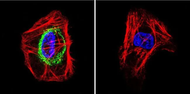
ICC/IF analysis of A431 cells using GTX25492 TSH receptor antibody [49]. Cells were probed without (right) or with(left) an antibody. Green : Primary antibody Blue : Nuclei Red : Actin Fixation : formaldehyde Dilution : 1:200 overnight at 4oC
![ICC/IF analysis of A431 cells using GTX25492 TSH receptor antibody [49]. Panel e is a no primary antibody control. Green : Primary antibody Blue : Nuclei Red : Actin Fixation : 4% paraformaldehyde Permeabilization : 0.1% Triton X-100 for 10 minute Dilution : 1:250 dilution in 0.1% BSA and incubated for 3 hours at room temperature](https://www.genetex.com/upload/website/prouct_img/normal/GTX25492/GTX25492_678_ICC-IF_w_23060722_951.webp "ICC/IF analysis of A431 cells using GTX25492 TSH receptor antibody [49]. Panel e is a no primary antibody control. Green : Primary antibody Blue : Nuclei Red : Actin Fixation : 4% paraformaldehyde Permeabilization : 0.1% Triton X-100 for 10 minute Dilution : 1:250 dilution in 0.1% BSA and incubated for 3 hours at room temperature")
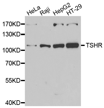
![WB analysis of membrane-enriched extracts (30 μg lysate) of A-431 (Lane 1), IMR-32 (Lane 2) and Jurkat (Lane 3) using GTX25492 TSH receptor antibody [49]. Dilution : 1:250](https://www.genetex.com/upload/website/prouct_img/normal/GTX25492/GTX25492_1787_WB_w_23060722_802.webp "WB analysis of membrane-enriched extracts (30 μg lysate) of A-431 (Lane 1), IMR-32 (Lane 2) and Jurkat (Lane 3) using GTX25492 TSH receptor antibody [49]. Dilution : 1:250")
ICC/IF analysis of A431 cells using GTX25492 TSH receptor antibody [49]. Cells were probed without (right) or with(left) an antibody. Green : Primary antibody Blue : Nuclei Red : Actin Fixation : formaldehyde Dilution : 1:200 overnight at 4oC
TSH receptor antibody [49]
GTX25492
ApplicationsImmunoFluorescence, Western Blot, ELISA, ImmunoCytoChemistry
Product group Antibodies
ReactivityHuman, Porcine
TargetTSHR
Overview
- SupplierGeneTex
- Product NameTSH receptor antibody [49]
- Delivery Days Customer9
- Application Supplier NoteWB: 1:250. ICC/IF: 1:250. *Optimal dilutions/concentrations should be determined by the researcher.Not tested in other applications.
- ApplicationsImmunoFluorescence, Western Blot, ELISA, ImmunoCytoChemistry
- CertificationResearch Use Only
- ClonalityMonoclonal
- Clone ID49
- ConjugateUnconjugated
- Gene ID7253
- Target nameTSHR
- Target descriptionthyroid stimulating hormone receptor
- Target synonymsCHNG1, LGR3, hTSHR-I, thyrotropin receptor, TSH receptor, seven transmembrane helix receptor, thyrotropin receptor-I, hTSHR-I
- HostMouse
- IsotypeIgG1
- Protein IDP16473
- Protein NameThyrotropin receptor
- Scientific DescriptionThe protein encoded by this gene is a membrane protein and a major controller of thyroid cell metabolism. The encoded protein is a receptor for thyrothropin and thyrostimulin, and its activity is mediated by adenylate cyclase. Defects in this gene are a cause of several types of hyperthyroidism. Three transcript variants encoding different isoforms have been found for this gene. [provided by RefSeq, Dec 2008]
- ReactivityHuman, Porcine
- Storage Instruction-20°C or -80°C,2°C to 8°C
- UNSPSC41116161
Datasheet
Related products
Product group Antibodies
Anti-TSHR AntibodyA44129
ApplicationsWestern Blot
ReactivityHuman, Mouse, Rat
- SizePrice
Product group Antibodies
Anti-TSHR Antibody144-06781
ApplicationsImmunoFluorescence, Western Blot, ImmunoHistoChemistry
ReactivityHuman, Mouse, Rat
TargetTSHR
- SizePrice

Product group Antibodies
Anti-TSH Receptor/TSHR Antibody Picoband(r)A00576-1-CARRIER-FREE
ApplicationsWestern Blot
ReactivityHuman, Mouse, Rat
TargetTSHR
- SizePrice
Product group Antibodies
TSHR Polyclonal AntibodyBS-0206R
ApplicationsFlow Cytometry, ImmunoFluorescence, ELISA, ImmunoCytoChemistry, ImmunoHistoChemistry, ImmunoHistoChemistry Frozen, ImmunoHistoChemistry Paraffin
ReactivityBovine, Chicken, Equine, Guinea Pig, Human, Porcine, Rabbit, Rat
TargetTSHR
- SizePrice
Product group Antibodies
TSHR AntibodyCSB-PA11189A0RB
ApplicationsImmunoFluorescence, ELISA
ReactivityHuman
TargetTSHR
- SizePrice
Product group Antibodies

Goat anti-TSHR (aa101-115)EB11115
ApplicationsWestern Blot, ELISA
ReactivityBovine, Human
TargetTSHR
- SizePrice
Product group Antibodies
Tshr Polyclonal AntibodyCAC10411
ApplicationsImmunoFluorescence, ELISA
TargetTSHR
- SizePrice
Product group Antibodies

TSH Receptor / TSHR AntibodyLS-C401111
ApplicationsELISA, ImmunoHistoChemistry
ReactivityHuman
TargetTSHR
- SizePrice
Product group Antibodies
TSH receptor antibodyGTX71175
ApplicationsImmunoHistoChemistry, ImmunoHistoChemistry Paraffin
ReactivityHuman, Monkey
TargetTSHR
- SizePrice