

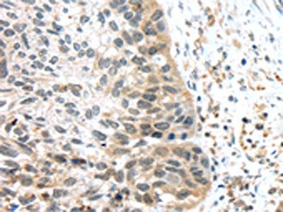
The image on the left is immunohistochemistry of paraffin-embedded Human esophagus cancer tissue using CSB-PA696394(TSPY1/TSPY3 Antibody) at dilution 1/45, on the right is treated with synthetic peptide. (Original magnification: x200)
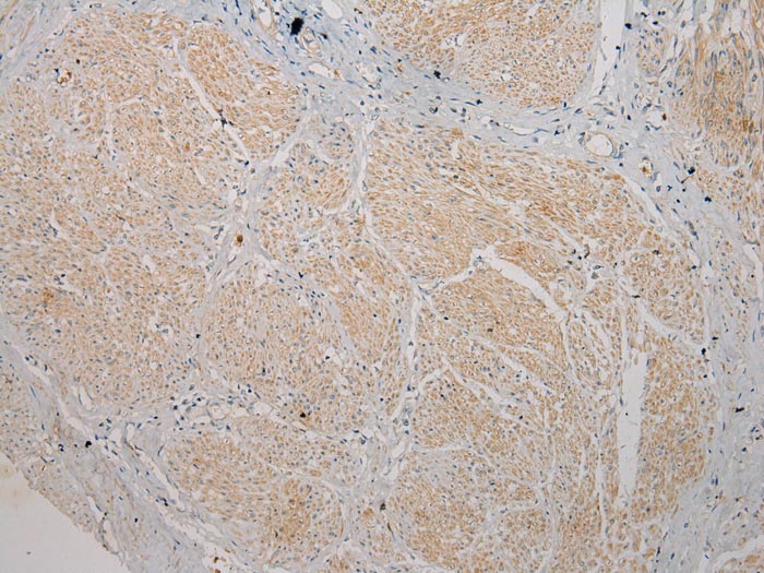
The image on the left is immunohistochemistry of paraffin-embedded Human esophagus cancer tissue using CSB-PA696394(TSPY1/TSPY3 Antibody) at dilution 1/45, on the right is treated with synthetic peptide. (Original magnification: x200)
TSPY1/TSPY3 Antibody
CSB-PA696394
ApplicationsELISA, ImmunoHistoChemistry
Product group Antibodies
ReactivityHuman
TargetTSPY1
Overview
- SupplierCusabio
- Product NameTSPY1/TSPY3 Antibody
- Delivery Days Customer20
- ApplicationsELISA, ImmunoHistoChemistry
- CertificationResearch Use Only
- ClonalityPolyclonal
- ConjugateUnconjugated
- Gene ID7258
- Target nameTSPY1
- Target descriptiontestis specific protein Y-linked 1
- Target synonymsCT78, DYS14, TSPY, pJA923, testis-specific Y-encoded protein 1, cancer/testis antigen 78, testis specific protein, Y-linked
- HostRabbit
- IsotypeIgG
- Protein IDP0CV98
- Protein NameTestis-specific Y-encoded protein 3
- Scientific DescriptionThe protein encoded by this gene is found only in testicular tissue and may be involved in spermatogenesis. Approximately 35 copies of this gene are present in humans, but only a single, nonfunctional orthologous gene is found in mouse. Two transcript variants encoding different isoforms have been found for this gene.
- ReactivityHuman
- Storage Instruction-20°C or -80°C
- UNSPSC41116161
Related products
Product group Antibodies
Anti-TSPY2 (Center) Antibody102-23936
ApplicationsWestern Blot
TargetTSPY1
- SizePrice

Product group Antibodies
Anti-TSPY1/2/3/4 Antibody Picoband(r)A04215-1-CARRIER-FREE
ApplicationsFlow Cytometry, Western Blot, ELISA
ReactivityHuman
TargetTSPY1
- SizePrice
Product group Antibodies
TSPY1 Polyclonal AntibodyBS-6607R
ApplicationsImmunoFluorescence, Western Blot, ELISA, ImmunoCytoChemistry, ImmunoHistoChemistry, ImmunoHistoChemistry Frozen, ImmunoHistoChemistry Paraffin
ReactivityHuman
TargetTSPY1
- SizePrice
Product group Antibodies
Anti-TSPY1 AntibodyA46354
ApplicationsImmunoHistoChemistry
ReactivityHuman
- SizePrice
Product group Antibodies
TSPY1/TSPY3 AntibodyPACO20781
ApplicationsELISA, ImmunoHistoChemistry
ReactivityHuman
TargetTSPY1
- SizePrice

Product group Antibodies
Anti-TSPY1 AntibodyHPA067289
ApplicationsImmunoCytoChemistry
ReactivityHuman
TargetTSPY1
- SizePrice
Product group Antibodies

TSPY1 / TSPY AntibodyLS-C771477
ApplicationsELISA, ImmunoHistoChemistry
ReactivityHuman
TargetTSPY1
- SizePrice