

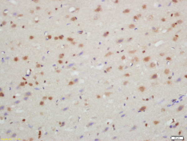
Formalin-fixed and paraffin embedded rat brain labeled with Rabbit Anti-TXNIP Polyclonal Antibody, Unconjugated (bs-3897R) at 1:200 followed by conjugation to the secondary antibody and DAB staining\n
Formalin-fixed and paraffin embedded rat brain labeled with Rabbit Anti-TXNIP Polyclonal Antibody, Unconjugated (bs-3897R) at 1:200 followed by conjugation to the secondary antibody and DAB staining\n
TXNIP Polyclonal Antibody
BS-3897R
ApplicationsFlow Cytometry, ImmunoFluorescence, Western Blot, ELISA, ImmunoCytoChemistry, ImmunoHistoChemistry, ImmunoHistoChemistry Frozen, ImmunoHistoChemistry Paraffin
Product group Antibodies
ReactivityCanine, Human, Mouse, Porcine, Rabbit, Rat
TargetTXNIP
Overview
- SupplierBioss
- Product NameTXNIP Polyclonal Antibody
- Delivery Days Customer16
- ApplicationsFlow Cytometry, ImmunoFluorescence, Western Blot, ELISA, ImmunoCytoChemistry, ImmunoHistoChemistry, ImmunoHistoChemistry Frozen, ImmunoHistoChemistry Paraffin
- Applications SupplierWB(1:300-5000), ELISA(1:500-1000), FCM(1:20-100), IHC-P(1:200-400), IHC-F(1:100-500), IF(IHC-P)(1:50-200), IF(IHC-F)(1:50-200), IF(ICC)(1:50-200)
- CertificationResearch Use Only
- ClonalityPolyclonal
- Concentration1 ug/ul
- ConjugateUnconjugated
- Gene ID10628
- Target nameTXNIP
- Target descriptionthioredoxin interacting protein
- Target synonymsARRDC6, EST01027, HHCPA78, THIF, VDUP1, thioredoxin-interacting protein, thioredoxin binding protein 2, upregulated by 1,25-dihydroxyvitamin D-3, vitamin D3 up-regulated protein 1
- HostRabbit
- IsotypeIgG
- Protein IDQ9H3M7
- Protein NameThioredoxin-interacting protein
- ReactivityCanine, Human, Mouse, Porcine, Rabbit, Rat
- Storage Instruction-20°C
- UNSPSC41116161
References
- Silymarin attenuated paraquat-induced cytotoxicity in macrophage by regulating Trx/TXNIP complex, inhibiting NLRP3 inflammasome activation and apoptosis. Liu Z et al., 2018 Feb, Toxicol In VitroRead this paper
Datasheet
Related products
Product group Antibodies
Anti-TXNIP AntibodyA81009
ApplicationsImmunoFluorescence, Western Blot, ImmunoCytoChemistry
ReactivityHuman, Mouse, Rat
- SizePrice
Product group Antibodies
Anti-TXNIP Antibody144-66751
ApplicationsWestern Blot
ReactivityHuman, Mouse
TargetTXNIP
- SizePrice
Product group Antibodies

TXNIP AntibodyLS-C771531
ApplicationsELISA, ImmunoHistoChemistry
ReactivityHuman, Mouse, Rat
TargetTXNIP
- SizePrice

Product group Antibodies
Anti-TXNIP Antibody Picoband(r)A01409-1-CARRIER-FREE
ApplicationsFlow Cytometry, Western Blot, ELISA, ImmunoHistoChemistry
ReactivityHuman, Mouse, Rat
TargetTXNIP
- SizePrice
Product group Antibodies
TXNIP AntibodyCSB-PA296101
ApplicationsELISA, ImmunoHistoChemistry
ReactivityHuman, Mouse, Rat
TargetTXNIP
- SizePrice
Product group Antibodies
Txnip Polyclonal AntibodyCAC11357
ApplicationsImmunoFluorescence, ELISA
TargetTXNIP
- SizePrice
Product group Antibodies
TXNIP antibodyGTX31592
ApplicationsWestern Blot, ELISA, ImmunoHistoChemistry, ImmunoHistoChemistry Paraffin
ReactivityHuman, Mouse, Rat
TargetTXNIP
- SizePrice

Product group Antibodies
Anti-TXNIP AntibodyHPA031085
ApplicationsWestern Blot, ImmunoHistoChemistry
ReactivityHuman
TargetTXNIP
- SizePrice