

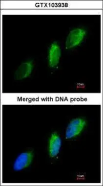
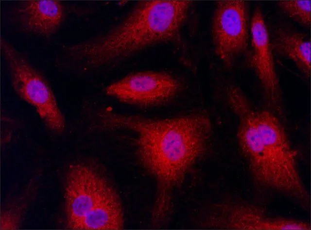
Immunofluorescence analysis of paraformaldehyde-fixed HeLa, using TYK2(GTX103938) antibody at 1:200 dilution.
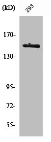
![Whole cell extract (30 μg) was separated by 5% SDS-PAGE, and the membrane was blotted with TYK2 antibody [C1C3] (GTX103938) diluted at 1:500. The HRP-conjugated anti-rabbit IgG antibody (GTX213110-01) was used to detect the primary antibody, and the signal was developed with Trident ECL plus-Enhanced.](https://www.genetex.com/upload/website/prouct_img/normal/GTX103938/GTX103938_39967_20210319_WB_2_w_23060120_664.webp "Whole cell extract (30 μg) was separated by 5% SDS-PAGE, and the membrane was blotted with TYK2 antibody [C1C3] (GTX103938) diluted at 1:500. The HRP-conjugated anti-rabbit IgG antibody (GTX213110-01) was used to detect the primary antibody, and the signal was developed with Trident ECL plus-Enhanced.")
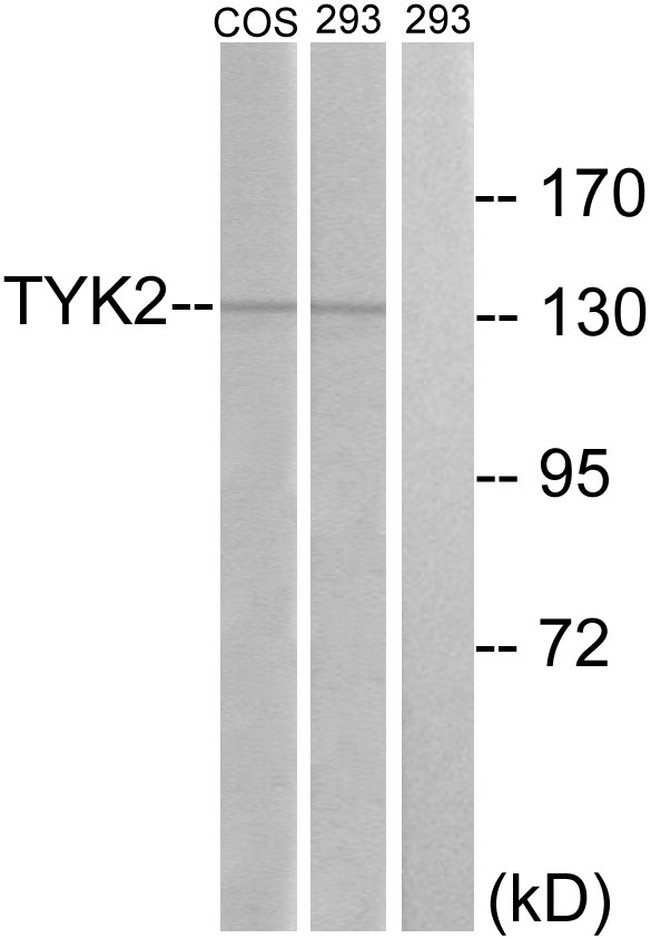
![Various whole cell extracts (30 μg) were separated by 5% SDS-PAGE, and the membrane was blotted with TYK2 antibody [C1C3] (GTX103938) diluted at 1:500. The HRP-conjugated anti-rabbit IgG antibody (GTX213110-01) was used to detect the primary antibody, and the signal was developed with Trident ECL plus-Enhanced.](https://www.genetex.com/upload/website/prouct_img/normal/GTX103938/GTX103938_39967_20210319_WB_w_23060120_853.webp "Various whole cell extracts (30 μg) were separated by 5% SDS-PAGE, and the membrane was blotted with TYK2 antibody [C1C3] (GTX103938) diluted at 1:500. The HRP-conjugated anti-rabbit IgG antibody (GTX213110-01) was used to detect the primary antibody, and the signal was developed with Trident ECL plus-Enhanced.")
Immunofluorescence analysis of paraformaldehyde-fixed HeLa, using TYK2(GTX103938) antibody at 1:200 dilution.
TYK2 antibody [C1C3]
GTX103938
ApplicationsImmunoFluorescence, Western Blot, ImmunoCytoChemistry
Product group Antibodies
ReactivityHuman
TargetTYK2
Overview
- SupplierGeneTex
- Product NameTYK2 antibody [C1C3]
- Delivery Days Customer9
- Application Supplier NoteWB: 1:500-1:3000. ICC/IF: 1:100-1:1000. *Optimal dilutions/concentrations should be determined by the researcher.Not tested in other applications.
- ApplicationsImmunoFluorescence, Western Blot, ImmunoCytoChemistry
- CertificationResearch Use Only
- ClonalityPolyclonal
- Concentration1 mg/ml
- ConjugateUnconjugated
- Gene ID7297
- Target nameTYK2
- Target descriptiontyrosine kinase 2
- Target synonymsIMD35, JTK1, non-receptor tyrosine-protein kinase TYK2
- HostRabbit
- IsotypeIgG
- Protein IDP29597
- Protein NameNon-receptor tyrosine-protein kinase TYK2
- Scientific DescriptionThis gene encodes a member of the tyrosine kinase and, more specifically, the Janus kinases (JAKs) protein families. This protein associates with the cytoplasmic domain of type I and type II cytokine receptors and promulgate cytokine signals by phosphorylating receptor subunits. It is also component of both the type I and type III interferon signaling pathways. As such, it may play a role in anti-viral immunity. A mutation in this gene has been associated with hyperimmunoglobulin E syndrome (HIES) - a primary immunodeficiency characterized by elevated serum immunoglobulin E. [provided by RefSeq]
- ReactivityHuman
- Storage Instruction-20°C or -80°C,2°C to 8°C
- UNSPSC41116161
Datasheet
Related products
Product group Antibodies
Anti-TYK2 AntibodyA96363
ApplicationsImmunoFluorescence, Western Blot, ELISA, ImmunoHistoChemistry
ReactivityHuman, Mouse
- SizePrice
Product group Antibodies
Anti-TYK2 Antibody144-65477
ApplicationsWestern Blot
ReactivityHuman, Mouse
TargetTYK2
- SizePrice

Product group Antibodies
Anti-TYK2 Antibody Picoband(r)A01642-3-CARRIER-FREE
ApplicationsFlow Cytometry, Western Blot, ELISA
ReactivityHuman
TargetTYK2
- SizePrice
Product group Antibodies
ApplicationsImmunoFluorescence, ImmunoCytoChemistry, ImmunoHistoChemistry, ImmunoHistoChemistry Frozen, ImmunoHistoChemistry Paraffin
ReactivityCanine, Human, Mouse, Rat
TargetTYK2
- SizePrice
Product group Antibodies
TYK2 AntibodyCSB-PA004352
ApplicationsImmunoFluorescence, Western Blot, ELISA, ImmunoHistoChemistry
ReactivityHuman, Monkey, Mouse
TargetTYK2
- SizePrice
Product group Antibodies
ApplicationsWestern Blot, ELISA, ImmunoCytoChemistry, ImmunoHistoChemistry, ImmunoHistoChemistry Frozen, ImmunoHistoChemistry Paraffin
TargetTYK2
- SizePrice
Product group Antibodies
TYK2 antibodyGTX30812
ApplicationsImmunoFluorescence, Western Blot, ImmunoCytoChemistry
ReactivityHuman
TargetTYK2
- SizePrice
Product group Antibodies
TYK2 antibodyGTX31515
ApplicationsImmunoFluorescence, Western Blot, ELISA, ImmunoCytoChemistry
ReactivityHuman, Mouse, Rat
TargetTYK2
- SizePrice
Product group Antibodies

TYK2 AntibodyLS-C331920
ApplicationsWestern Blot
ReactivityHuman, Mouse
TargetTYK2
- SizePrice

Product group Antibodies
Anti-TYK2 AntibodyHPA005157
ApplicationsWestern Blot, ImmunoCytoChemistry
ReactivityHuman
TargetTYK2
- SizePrice