

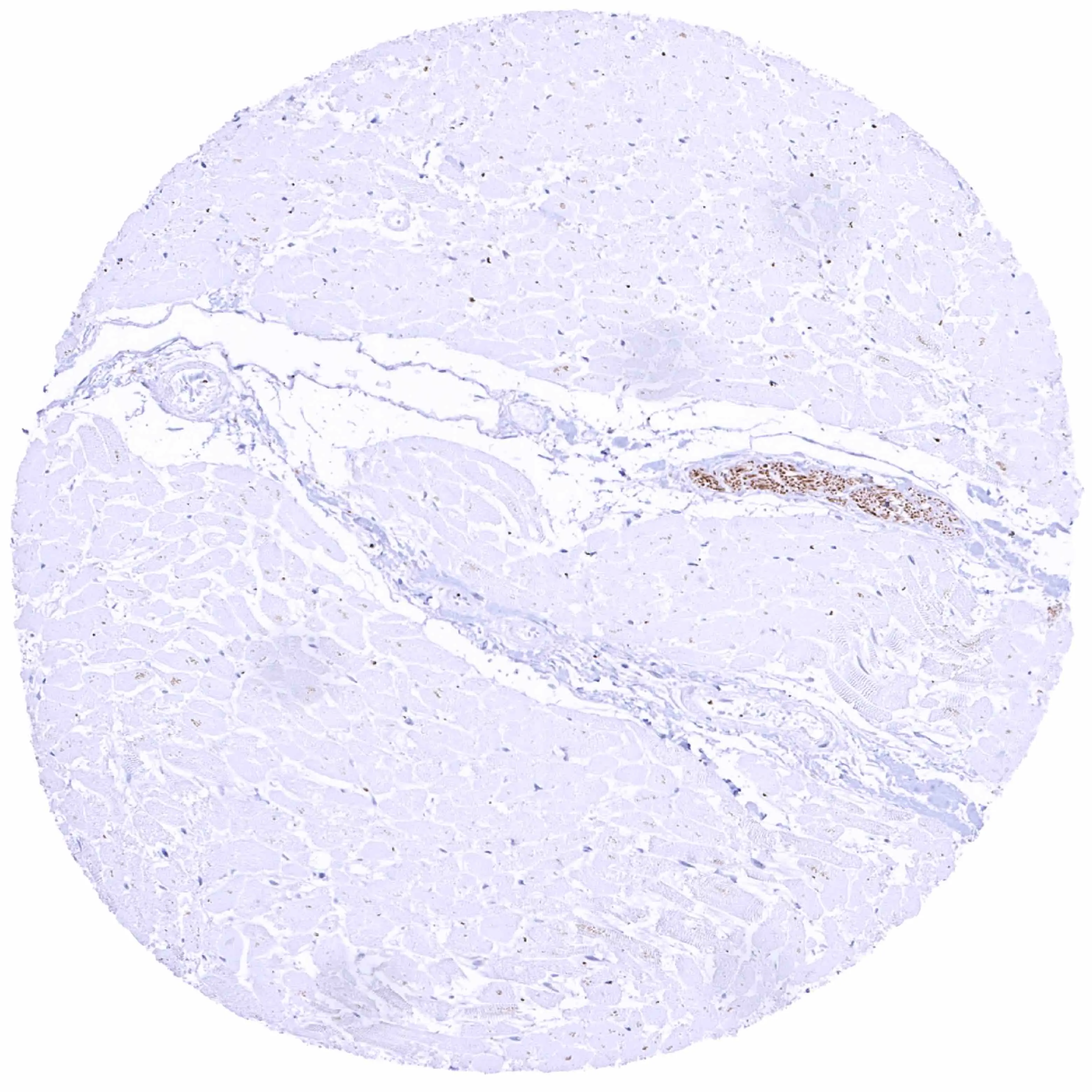
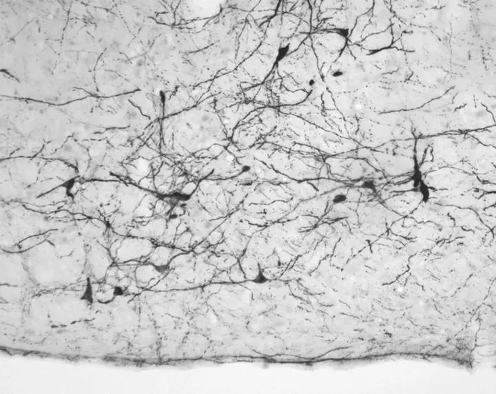
IHC-P analysis of human heart muscle tissue using GTX639936 Tyrosine Hydroxylase antibody [HMV312] HistoMAX?. Strong tyrosine hydroxylase staining of fibers in a small nerve.
![IHC-P analysis of human ovarian stroma tissue using GTX639936 Tyrosine Hydroxylase antibody [HMV312] HistoMAX?. A small nerve shows distinct tyrosine hydroxylase positivity of nerve fibers. Individual nerve fibers also show tyrosine hydroxylase staining.](https://www.genetex.com/upload/website/prouct_img/normal/GTX639936/GTX639936_20240408_IHC-P_1_24040801_341.webp "IHC-P analysis of human ovarian stroma tissue using GTX639936 Tyrosine Hydroxylase antibody [HMV312] HistoMAX?. A small nerve shows distinct tyrosine hydroxylase positivity of nerve fibers. Individual nerve fibers also show tyrosine hydroxylase staining.")
![IHC-P analysis of human paraganglioma tissue using GTX639936 Tyrosine Hydroxylase antibody [HMV312] HistoMAX?. Paraganglioma lacking tyrosine hydroxylase staining.](https://www.genetex.com/upload/website/prouct_img/normal/GTX639936/GTX639936_20250214_IHC-P_1_25021323_377.webp "IHC-P analysis of human paraganglioma tissue using GTX639936 Tyrosine Hydroxylase antibody [HMV312] HistoMAX?. Paraganglioma lacking tyrosine hydroxylase staining.")
![IHC-P analysis of human pheochromocytoma of the adrenal gland tissue using GTX639936 Tyrosine Hydroxylase antibody [HMV312] HistoMAX?. A strong tyrosine hydroxylase staining of all tumor cells.](https://www.genetex.com/upload/website/prouct_img/normal/GTX639936/GTX639936_20250214_IHC-P_2_25021323_601.webp "IHC-P analysis of human pheochromocytoma of the adrenal gland tissue using GTX639936 Tyrosine Hydroxylase antibody [HMV312] HistoMAX?. A strong tyrosine hydroxylase staining of all tumor cells.")
![IHC-P analysis of human adrenal gland tissue using GTX639936 Tyrosine Hydroxylase antibody [HMV312] HistoMAX?. A strong cytoplasmic and membranous tyrosine hydroxylase staining of medullary cells while adrenocortical cells remain negative.](https://www.genetex.com/upload/website/prouct_img/normal/GTX639936/GTX639936_20250214_IHC-P_25021323_417.webp "IHC-P analysis of human adrenal gland tissue using GTX639936 Tyrosine Hydroxylase antibody [HMV312] HistoMAX?. A strong cytoplasmic and membranous tyrosine hydroxylase staining of medullary cells while adrenocortical cells remain negative.")
IHC-P analysis of human heart muscle tissue using GTX639936 Tyrosine Hydroxylase antibody [HMV312] HistoMAX?. Strong tyrosine hydroxylase staining of fibers in a small nerve.
Tyrosine Hydroxylase antibody [HMV312] HistoMAX(tm)
GTX639936
ApplicationsImmunoHistoChemistry, ImmunoHistoChemistry Paraffin
Product group Antibodies
ReactivityHuman
TargetTH
Overview
- SupplierGeneTex
- Product NameTyrosine Hydroxylase antibody [HMV312] HistoMAX(tm)
- Delivery Days Customer7
- Application Supplier NoteIHC-P: 1:100-1:200. *Optimal dilutions/concentrations should be determined by the researcher.Not tested in other applications.
- ApplicationsImmunoHistoChemistry, ImmunoHistoChemistry Paraffin
- CertificationResearch Use Only
- ClonalityMonoclonal
- Clone IDHMV312
- Concentration7 ug/ml
- ConjugateUnconjugated
- Gene ID7054
- Target nameTH
- Target descriptiontyrosine hydroxylase
- Target synonymsDYT14, DYT5b, TYH, tyrosine 3-monooxygenase, dystonia 14, tyrosine 3-hydroxylase
- HostRabbit
- IsotypeIgG
- Protein IDP07101
- Protein NameTyrosine 3-monooxygenase
- Scientific DescriptionThe protein encoded by this gene is involved in the conversion of tyrosine to dopamine. It is the rate-limiting enzyme in the synthesis of catecholamines, hence plays a key role in the physiology of adrenergic neurons. Mutations in this gene have been associated with autosomal recessive Segawa syndrome. Alternatively spliced transcript variants encoding different isoforms have been noted for this gene. [provided by RefSeq, Jul 2008]
- ReactivityHuman
- Storage Instruction-20°C or -80°C,2°C to 8°C
- UNSPSC41116161
Datasheet
Related products
Product group Antibodies
ApplicationsFlow Cytometry, ImmunoFluorescence, Western Blot, ImmunoCytoChemistry, ImmunoHistoChemistry
ReactivityHuman, Mouse, Rat
- SizePrice
Product group Antibodies
Anti-TH Antibody144-00028
ApplicationsImmunoFluorescence, Western Blot, ImmunoHistoChemistry
ReactivityHuman, Mouse, Rat
TargetTH
- SizePrice
Product group Antibodies

ApplicationsImmunoFluorescence, Western Blot, ELISA, ImmunoCytoChemistry, ImmunoHistoChemistry
- SizePrice

Product group Antibodies
Anti-TH AntibodyAMAB91112
ApplicationsImmunoHistoChemistry
ReactivityHuman, Mouse, Rat
TargetTH
- SizePrice
Product group Antibodies
TH Polyclonal AntibodyCAC14884
ApplicationsWestern Blot, ELISA, ImmunoHistoChemistry
ReactivityMouse, Rat
TargetTH
- SizePrice
Product group Antibodies
TH AntibodyCSB-PA004284
ApplicationsImmunoFluorescence, Western Blot, ELISA, ImmunoHistoChemistry
ReactivityHuman, Mouse, Rat
TargetTH
- SizePrice
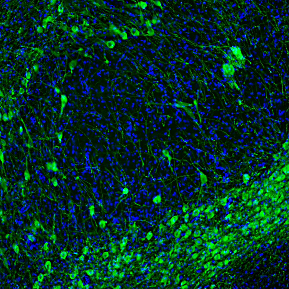
![Tyrosine Hydroxylase antibody [N1C1] detects Tyrosine Hydroxylase protein by immunohistochemical analysis. Sample: Frozen sectioned adult mouse retina. Green: Tyrosine Hydroxylase protein stained by Tyrosine Hydroxylase antibody [N1C1] (GTX102424) diluted at 1:250. Red: beta Tubulin 3/ TUJ1, stained by beta Tubulin 3/ TUJ1 antibody [GT11710] (GTX631836) diluted at 1:250. Blue: Fluoroshield with DAPI (GTX30920).](https://www.genetex.com/upload/website/prouct_img/normal/GTX102424/GTX102424_39974_20160830_IHC-Fr_w_23060100_342.webp)
Product group Antibodies
Tyrosine Hydroxylase antibody [N1C1]GTX102424
ApplicationsImmunoFluorescence, Western Blot, ImmunoCytoChemistry, ImmunoHistoChemistry, ImmunoHistoChemistry Frozen, ImmunoHistoChemistry Paraffin
ReactivityHuman, Mouse, Rat, Zebra Fish
TargetTH
- SizePrice
Product group Antibodies
Tyrosine Hydroxylase antibodyGTX113016
ApplicationsImmunoFluorescence, Western Blot, ImmunoCytoChemistry, ImmunoHistoChemistry, ImmunoHistoChemistry Frozen, ImmunoHistoChemistry Paraffin
ReactivityBovine, Human, Mammals, Mouse, Rat, Zebra Fish
TargetTH
- SizePrice
Product group Antibodies
Tyrosine Hydroxylase antibodyGTX85470
ApplicationsImmunoFluorescence, Western Blot, ImmunoCytoChemistry, ImmunoHistoChemistry, ImmunoHistoChemistry Paraffin
ReactivityHuman, Mouse, Rat
TargetTH
- SizePrice