

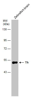
Zebrafish tissue extract (30 μg) was separated by 7.5% SDS-PAGE, and the membrane was blotted with Tyrosine Hydroxylase antibody (GTX113016) diluted at 1:500.
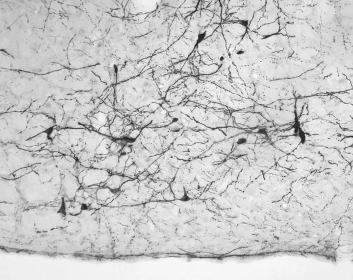
, using Tyrosine Hydroxylase(GTX113016) antibody at 1:100 dilution.
Antigen Retrieval: Citrate buffer, pH 6.0, 15 min")
![Tyrosine Hydroxylase antibody detects Tyrosine Hydroxylase protein by immunohistochemical analysis. Sample: Frozen sectioned adult mouse retina. Green: Tyrosine Hydroxylase protein stained by Tyrosine Hydroxylase antibody (GTX113016) diluted at 1:250. Red: beta Tubulin 3/ TUJ1, stained by beta Tubulin 3/ TUJ1 antibody [GT11710] (GTX631836) diluted at 1:250. Blue: Fluoroshield with DAPI (GTX30920).](https://www.genetex.com/upload/website/prouct_img/normal/GTX113016/GTX113016_42116_20160830_IHC-Fr_w_23060500_404.webp "Tyrosine Hydroxylase antibody detects Tyrosine Hydroxylase protein by immunohistochemical analysis. Sample: Frozen sectioned adult mouse retina. Green: Tyrosine Hydroxylase protein stained by Tyrosine Hydroxylase antibody (GTX113016) diluted at 1:250. Red: beta Tubulin 3/ TUJ1, stained by beta Tubulin 3/ TUJ1 antibody [GT11710] (GTX631836) diluted at 1:250. Blue: Fluoroshield with DAPI (GTX30920).")
, using Tyrosine Hydroxylase(GTX113016) antibody at 1:100 dilution.
Antigen Retrieval: Citrate buffer, pH 6.0, 15 min")
 diluted at 1:1000. Blue: Fluoroshield with DAPI (GTX30920). Antigen Retrieval: Citrate buffer, pH 6.0, 15 min")
 dilution: 1:500 The HRP-conjugated anti-rabbit IgG antibody (GTX213110-01) was used to detect the primary antibody.")
 diluted at 1:400. Red: beta Tubulin 3/ TUJ1 protein stained by beta Tubulin 3/ TUJ1 antibody (GTX631836) diluted at 1:200. Blue: Hoechst 33342 staining.")
 was separated by 7.5% SDS-PAGE, and the membrane was blotted with Tyrosine Hydroxylase antibody (GTX113016) diluted at 1:1000.")
 diluted at 1:2500.
Antigen Retrieval: Citrate buffer, pH 6.0, 15 min")
 were separated by 7.5% SDS-PAGE, and the membrane was blotted with Tyrosine Hydroxylase antibody (GTX113016) diluted at 1:500. The HRP-conjugated anti-rabbit IgG antibody (GTX213110-01) was used to detect the primary antibody.")
Zebrafish tissue extract (30 μg) was separated by 7.5% SDS-PAGE, and the membrane was blotted with Tyrosine Hydroxylase antibody (GTX113016) diluted at 1:500.
Tyrosine Hydroxylase antibody
GTX113016
ApplicationsImmunoFluorescence, Western Blot, ImmunoCytoChemistry, ImmunoHistoChemistry, ImmunoHistoChemistry Frozen, ImmunoHistoChemistry Paraffin
Product group Antibodies
ReactivityBovine, Human, Mammals, Mouse, Rat, Zebra Fish
TargetTH
Overview
- SupplierGeneTex
- Product NameTyrosine Hydroxylase antibody
- Delivery Days Customer9
- Application Supplier NoteWB: 1:500-1:3000. ICC/IF: 1:100-1:1000. IHC-P: 1:100-1:1000. IHC-Fr: 1:100-1:1000. *Optimal dilutions/concentrations should be determined by the researcher.Not tested in other applications.
- ApplicationsImmunoFluorescence, Western Blot, ImmunoCytoChemistry, ImmunoHistoChemistry, ImmunoHistoChemistry Frozen, ImmunoHistoChemistry Paraffin
- CertificationResearch Use Only
- ClonalityPolyclonal
- Concentration0.21 mg/ml
- ConjugateUnconjugated
- Gene ID7054
- Target nameTH
- Target descriptiontyrosine hydroxylase
- Target synonymsDYT14, DYT5b, TYH, tyrosine 3-monooxygenase, dystonia 14, tyrosine 3-hydroxylase
- HostRabbit
- IsotypeIgG
- Protein IDP07101
- Protein NameTyrosine 3-monooxygenase
- Scientific DescriptionThe protein encoded by this gene is involved in the conversion of tyrosine to dopamine. It is the rate-limiting enzyme in the synthesis of catecholamines, hence plays a key role in the physiology of adrenergic neurons. Mutations in this gene have been associated with autosomal recessive Segawa syndrome. Alternatively spliced transcript variants encoding different isoforms have been noted for this gene. [provided by RefSeq]
- ReactivityBovine, Human, Mammals, Mouse, Rat, Zebra Fish
- Storage Instruction-20°C or -80°C,2°C to 8°C
- UNSPSC12352203
References
- Wu CY, Song DF, Lu TH, et al. Klotho Null Mutation Indirectly Leads to Age-Related Lacrimal Gland Degeneration in Mutant Mice. Biology (Basel). 2023,12(10). doi: 10.3390/biology12101328Read this paper
- Ikeda N, Kawasaki M, Baba K, et al. Chemogenetic Activation of Oxytocin Neurons Improves Pain in a Reserpine-induced Fibromyalgia Rat Model. Neuroscience. 2023,528:37-53. doi: 10.1016/j.neuroscience.2023.07.028Read this paper
- Yamamoto S, Matsui A, Ohyagi M, et al. In Vitro Generation of Brain Regulatory T Cells by Co-culturing With Astrocytes. Front Immunol. 2022,13:960036. doi: 10.3389/fimmu.2022.960036Read this paper
- Reimer L, Haikal C, Gram H, et al. Low dose DMSO treatment induces oligomerization and accelerates aggregation of α-synuclein. Sci Rep. 2022,12(1):3737. doi: 10.1038/s41598-022-07706-2Read this paper
- Song Y, Chu R, Cao F, et al. Dopaminergic Neurons in the Ventral Tegmental-Prelimbic Pathway Promote the Emergence of Rats from Sevoflurane Anesthesia. Neurosci Bull. 2022,38(4):417-428. doi: 10.1007/s12264-021-00809-2Read this paper
- Di Nisio A, Pannella M, Vogiatzis S, et al. Impairment of human dopaminergic neurons at different developmental stages by perfluoro-octanoic acid (PFOA) and differential human brain areas accumulation of perfluoroalkyl chemicals. Environ Int. 2022,158:106982. doi: 10.1016/j.envint.2021.106982Read this paper
- Fede C, Petrelli L, Guidolin D, et al. Evidence of a new hidden neural network into deep fasciae. Sci Rep. 2021,11(1):12623. doi: 10.1038/s41598-021-92194-zRead this paper
- El Manaa W, Duplan E, Goiran T, et al. Transcription- and phosphorylation-dependent control of a functional interplay between XBP1s and PINK1 governs mitophagy and potentially impacts Parkinson disease pathophysiology. Autophagy. 2021,17(12):4363-4385. doi: 10.1080/15548627.2021.1917129Read this paper
- Kawabe M, Hayashi A, Komatsu M, et al. Ninjinyoeito improves anxiety behavior in neuropeptide Y deficient zebrafish. Neuropeptides. 2021,87:102136. doi: 10.1016/j.npep.2021.102136Read this paper
- Wu J, Yu X, Xue K, et al. The Inhibition of miR-873 Provides Therapeutic Benefit in a Lipopolysaccharide-Induced Neuroinflammatory Model of Parkinson's Disease. Oxid Med Cell Longev. 2020,2020:8735249. doi: 10.1155/2020/8735249Read this paper
Datasheet
Related products
Product group Antibodies

ApplicationsImmunoFluorescence, Western Blot, ELISA, ImmunoCytoChemistry, ImmunoHistoChemistry
- SizePrice
Product group Antibodies
Anti-TH Antibody144-00028
ApplicationsImmunoFluorescence, Western Blot, ImmunoHistoChemistry
ReactivityHuman, Mouse, Rat
TargetTH
- SizePrice

Product group Antibodies
Anti-TH AntibodyAMAB91112
ApplicationsImmunoHistoChemistry
ReactivityHuman, Mouse, Rat
TargetTH
- SizePrice
![Tyrosine Hydroxylase antibody [N1C1] detects Tyrosine Hydroxylase protein by immunohistochemical analysis. Sample: Frozen sectioned adult mouse retina. Green: Tyrosine Hydroxylase protein stained by Tyrosine Hydroxylase antibody [N1C1] (GTX102424) diluted at 1:250. Red: beta Tubulin 3/ TUJ1, stained by beta Tubulin 3/ TUJ1 antibody [GT11710] (GTX631836) diluted at 1:250. Blue: Fluoroshield with DAPI (GTX30920).](https://www.genetex.com/upload/website/prouct_img/normal/GTX102424/GTX102424_39974_20160830_IHC-Fr_w_23060100_342.webp)
Product group Antibodies
References
Tyrosine Hydroxylase antibody [N1C1]GTX102424
ApplicationsImmunoFluorescence, Western Blot, ImmunoCytoChemistry, ImmunoHistoChemistry, ImmunoHistoChemistry Frozen, ImmunoHistoChemistry Paraffin
ReactivityHuman, Mouse, Rat, Zebra Fish
TargetTH
- SizePrice
![Mouse tissue extract (30 μg) was separated by 10% SDS-PAGE, and the membrane was blotted with Tyrosine Hydroxylase antibody [GT234] (GTX634481) diluted at 1:1000. The HRP-conjugated anti-mouse IgG antibody (GTX213111-01) was used to detect the primary antibody.](https://www.genetex.com/upload/website/prouct_img/normal/GTX634481/GTX634481_43143_20180518_WB_M_brain_w_23061202_591.webp)
Product group Antibodies
References
ApplicationsImmunoFluorescence, Western Blot, ImmunoCytoChemistry, ImmunoHistoChemistry, ImmunoHistoChemistry Paraffin
ReactivityHuman, Mouse, Rat
TargetTH
- SizePrice
![Whole cell extract (30 μg) was separated by 10% SDS-PAGE, and the membrane was blotted with Tyrosine Hydroxylase antibody [HL1762] (GTX637412) diluted at 1:1000. The HRP-conjugated anti-rabbit IgG antibody (GTX213110-01) was used to detect the primary antibody.](https://www.genetex.com/upload/website/prouct_img/normal/GTX637412/GTX637412_T-44795_20220908_WB_R_22091323_117.webp)
Product group Antibodies
ApplicationsWestern Blot, ImmunoHistoChemistry, ImmunoHistoChemistry Paraffin
ReactivityHuman, Mouse, Rat
TargetTH
- SizePrice
Product group Antibodies
Tyrosine Hydroxylase antibodyGTX85470
ApplicationsImmunoFluorescence, Western Blot, ImmunoCytoChemistry, ImmunoHistoChemistry, ImmunoHistoChemistry Paraffin
ReactivityHuman, Mouse, Rat
TargetTH
- SizePrice
Product group Antibodies
TH Polyclonal AntibodyCAC14884
ApplicationsWestern Blot, ELISA, ImmunoHistoChemistry
ReactivityMouse, Rat
TargetTH
- SizePrice