

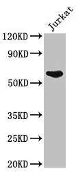
Western Blot Positive WB detected in: Jurkat whole cell lysate All lanes: UAP1 antibody at 2.5microg/ml Secondary Goat polyclonal to rabbit IgG at 1/50000 dilution Predicted band size: 59, 58 kDa Observed band size: 59 kDa

")
Western Blot Positive WB detected in: Jurkat whole cell lysate All lanes: UAP1 antibody at 2.5microg/ml Secondary Goat polyclonal to rabbit IgG at 1/50000 dilution Predicted band size: 59, 58 kDa Observed band size: 59 kDa
UAP1 Antibody
CSB-PA618067LA01HU
ApplicationsImmunoFluorescence, Western Blot, ELISA, ImmunoHistoChemistry
Product group Antibodies
ReactivityHuman
TargetUAP1
Overview
- SupplierCusabio
- Product NameUAP1 Antibody
- Delivery Days Customer20
- ApplicationsImmunoFluorescence, Western Blot, ELISA, ImmunoHistoChemistry
- CertificationResearch Use Only
- ClonalityPolyclonal
- ConjugateUnconjugated
- Gene ID6675
- Target nameUAP1
- Target descriptionUDP-N-acetylglucosamine pyrophosphorylase 1
- Target synonymsAGX, AGX1, AGX2, SPAG2, UDP-N-acetylhexosamine pyrophosphorylase, UDP-N-acetylgalactosamine pyrophosphorylase, UDP-N-acetylglucosamine diphosphorylase 1, UDP-N-acetylhexosamine pyrophosphorylase 1, antigen X, protein-pyrophosphorylation enzyme, sperm associated antigen 2, testis tissue sperm-binding protein Li 37a
- HostRabbit
- IsotypeIgG
- Protein IDQ16222
- Protein NameUDP-N-acetylhexosamine pyrophosphorylase
- Scientific DescriptionConverts UTP and GlcNAc-1-P into UDP-GlcNAc, and UTP and GalNAc-1-P into UDP-GalNAc. Isoform AGX1 has 2 to 3 times higher activity towards GalNAc-1-P, while isoform AGX2 has 8 times more activity towards GlcNAc-1-P.
- ReactivityHuman
- Storage Instruction-20°C or -80°C
- UNSPSC41116161
Related products
Product group Antibodies
Anti-UAP1 AntibodyA44037
ApplicationsWestern Blot
ReactivityHuman, Mouse, Rat
- SizePrice

Product group Antibodies
ApplicationsFlow Cytometry, ImmunoFluorescence, ImmunoPrecipitation, Western Blot, ImmunoCytoChemistry, ImmunoHistoChemistry
ReactivityHuman, Mouse, Rat
TargetUAP1
- SizePrice

Product group Antibodies
Anti-UAP1 AntibodyHPA014659
ApplicationsWestern Blot, ImmunoCytoChemistry, ImmunoHistoChemistry
ReactivityHuman
TargetUAP1
- SizePrice
Product group Antibodies

UAP1 AntibodyLS-C331908
ApplicationsWestern Blot, ImmunoHistoChemistry
ReactivityHuman, Mouse, Rat
TargetUAP1
- SizePrice
Product group Antibodies
UAP1 Polyclonal AntibodyCAC14780
ApplicationsImmunoFluorescence, Western Blot, ELISA, ImmunoHistoChemistry
TargetUAP1
- SizePrice
![UAP1 antibody [N1C3] detects UAP1 protein at cytoplasm by immunofluorescent analysis. Sample: HeLa cells were fixed in 4% paraformaldehyde at RT for 15 min. Green: UAP1 protein stained by UAP1 antibody [N1C3] (GTX103592) diluted at 1:200. Red: alpha Tubulin, a cytoskeleton marker, stained by alpha Tubulin antibody [B-5-1-2] (GTX11304) diluted at 1:10000. Blue: Hoechst 33342 staining.](https://www.genetex.com/upload/website/prouct_img/normal/GTX103592/GTX103592_39995_20150410_IFA_w_23060119_314.webp)
Product group Antibodies
UAP1 antibody [N1C3]GTX103592
ApplicationsImmunoFluorescence, Western Blot, ImmunoCytoChemistry, ImmunoHistoChemistry, ImmunoHistoChemistry Paraffin
ReactivityHuman
TargetUAP1
- SizePrice
Product group Antibodies
UAP1 Recombinant Antibody, AbBy Fluor-647 ConjugatedBSM-61991R-BF647
ApplicationsImmunoFluorescence, Western Blot
ReactivityHuman, Mouse, Rat
TargetUAP1
- SizePrice
Product group Antibodies
Anti-UAP1 (C-term) Antibody102-22902
ApplicationsWestern Blot
TargetUAP1
- SizePrice