

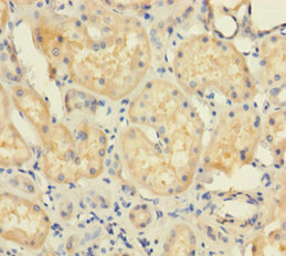
Immunohistochemistry of paraffin-embedded human kidney tissue using CSB-PA025435LA01HU at dilution of 1:100
")
Immunohistochemistry of paraffin-embedded human kidney tissue using CSB-PA025435LA01HU at dilution of 1:100
UBD Antibody
CSB-PA025435LA01HU
ApplicationsImmunoFluorescence, ELISA, ImmunoHistoChemistry
Product group Antibodies
ReactivityHuman
TargetUBD
Overview
- SupplierCusabio
- Product NameUBD Antibody
- Delivery Days Customer20
- ApplicationsImmunoFluorescence, ELISA, ImmunoHistoChemistry
- CertificationResearch Use Only
- ClonalityPolyclonal
- ConjugateUnconjugated
- Gene ID10537
- Target nameUBD
- Target descriptionubiquitin like modifier D
- Target synonymsFAT10, UBD-3, ubiquitin D, HLA-F adjacent transcript 10, diubiquitin, ubiquitin-like protein FAT10
- HostRabbit
- IsotypeIgG
- Protein IDO15205
- Protein NameUbiquitin D
- Scientific DescriptionUbiquitin-like protein modifier which can be covalently attached to target protein and subsequently leads to their degradation by the 26S proteasome, in a NUB1L-dependent manner. Probably functions as a survival factor. Conjugation ability activated by UBA6. Promotes the expression of the proteasome subunit beta type-9 (PSMB9/LMP2). Regulates TNF-alpha-induced and LPS-mediated activation of the central mediator of innate immunity NF-kappa-B by promoting TNF-alpha-mediated proteasomal degradation of ubiquitinated-I-kappa-B-alpha. Required for TNF-alpha-induced p65 nuclear translocation in renal tubular epithelial cells (RTECs). May be involved in dendritic cell (DC) maturation, the process by which immature dendritic cells differentiate into fully competent antigen-presenting cells that initiate T-cell responses. Mediates mitotic non-disjunction and chromosome instability, in long-term in vitro culture and cancers, by abbreviating mitotic phase and impairing the kinetochore localization of MAD2L1 during the prometaphase stage of the cell cycle. May be involved in the formation of aggresomes when proteasome is saturated or impaired. Mediates apoptosis in a caspase-dependent manner, especially in renal epithelium and tubular cells during renal diseases such as polycystic kidney disease and Human immunodeficiency virus (HIV)-associated nephropathy (HIVAN).
- ReactivityHuman
- Storage Instruction-20°C or -80°C
- UNSPSC41116161
Related products
Product group Antibodies
Anti-UBD AntibodyA30802
ApplicationsImmunoFluorescence, Western Blot, ImmunoHistoChemistry
ReactivityHuman, Mouse
- SizePrice
Product group Antibodies
Anti-UBD Antibody144-05491
ApplicationsImmunoFluorescence, Western Blot
ReactivityHuman, Mouse
TargetUBD
- SizePrice
Product group Antibodies

FAT10 / UBD AntibodyLS-C830673
ApplicationsELISA, ImmunoHistoChemistry
ReactivityHuman
TargetUBD
- SizePrice

Product group Antibodies
Anti-FAT10/UBD Picoband(r) AntibodyA01970-1-CARRIER-FREE
ApplicationsWestern Blot, ELISA
ReactivityHuman
TargetUBD
- SizePrice
Product group Antibodies
Ubiquitin D Recombinant Antibody, Biotin ConjugatedBSM-61330R-BIOTIN
ApplicationsWestern Blot, ImmunoHistoChemistry, ImmunoHistoChemistry Frozen, ImmunoHistoChemistry Paraffin
TargetUBD
- SizePrice
Product group Antibodies
ApplicationsImmunoPrecipitation, Western Blot, ImmunoCytoChemistry, ImmunoHistoChemistry
ReactivityMouse, Rat
TargetUBD
- SizePrice

Product group Antibodies
Anti-UBD-25ulHPA043710
ApplicationsWestern Blot, ImmunoCytoChemistry
ReactivityHuman
- SizePrice
Product group Antibodies
FAT10 antibodyGTX33566
ApplicationsImmunoFluorescence, Western Blot, ImmunoCytoChemistry
ReactivityHuman, Mouse
TargetUBD
- SizePrice