

UGT1A1 antibody
70R-1876
Product group Antibodies
Overview
- SupplierBiosynth
- Product NameUGT1A1 antibody
- Delivery Days Customer11
- CertificationResearch Use Only
- UNSPSC41116161
Related products
Product group Antibodies
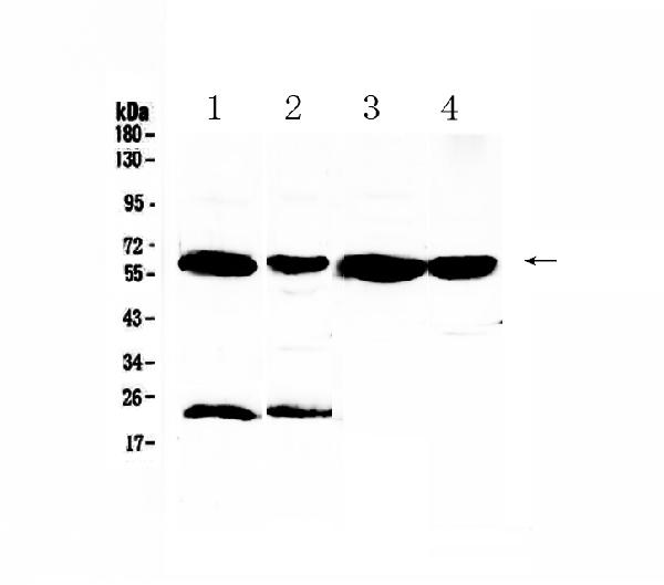
Anti-UGT1A1 AntibodyA31139
ApplicationsImmunoFluorescence, Western Blot, ImmunoHistoChemistry
ReactivityHuman
- SizePrice
Product group Antibodies
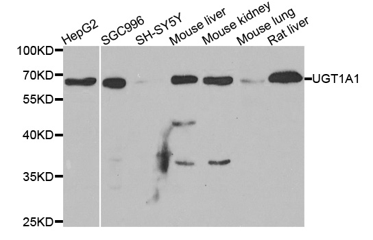
Anti-UGT1A1 Antibody144-01359
ApplicationsWestern Blot
ReactivityHuman, Mouse, Rat
TargetUGT1A1
- SizePrice
Product group Antibodies
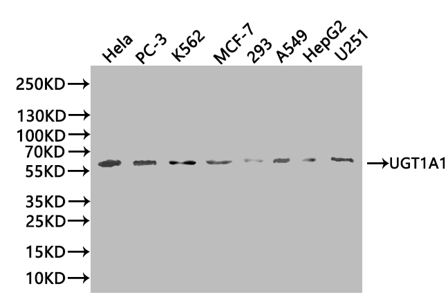
Anti-UGT1A1 Antibody Picoband(r)A01865-1-CARRIER-FREE
ApplicationsWestern Blot, ELISA, ImmunoHistoChemistry
ReactivityHuman, Mouse, Rat
TargetUGT1A1
- SizePrice
Product group Antibodies
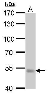
Ugt1A1 Polyclonal AntibodyCAC11384
ApplicationsWestern Blot, ELISA, ImmunoHistoChemistry
TargetUGT1A1
- SizePrice
Product group Antibodies
UGT1A1 AntibodyCSB-PA025570LA01HU
ApplicationsImmunoFluorescence, Western Blot, ELISA, ImmunoHistoChemistry
ReactivityHuman
TargetUGT1A1
- SizePrice
Product group Antibodies

UGT1A / UGT1A1 AntibodyLS-C334543
ApplicationsImmunoFluorescence, Western Blot, ImmunoHistoChemistry
ReactivityHuman
TargetUGT1A1
- SizePrice
Product group Antibodies
UGT1A antibodyGTX114131
ApplicationsWestern Blot, ImmunoHistoChemistry, ImmunoHistoChemistry Paraffin
ReactivityHuman, Mouse, Rat
TargetUGT1A1
- SizePrice
Product group Antibodies
Anti-PHPT1 AntibodyCAB13590
ApplicationsWestern Blot, ELISA
ReactivityHuman
TargetUGT1A1
- SizePrice