

V-ATPase B1 antibody [N1C1-2]
GTX110341
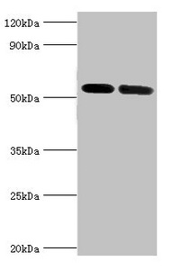
ApplicationsWestern Blot
Product group Antibodies
ReactivityHuman
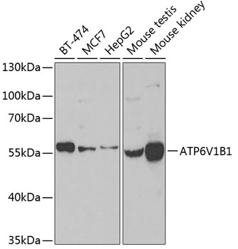
TargetATP6V1B1
Overview
- SupplierGeneTex
- Product NameV-ATPase B1 antibody [N1C1-2]
- Delivery Days Customer9
- ApplicationsWestern Blot
- CertificationResearch Use Only
- ClonalityPolyclonal
- Concentration0.42 mg/ml
- ConjugateUnconjugated
- Gene ID525
- Target nameATP6V1B1
- Target descriptionATPase H+ transporting V1 subunit B1
- Target synonymsATP6B1, DRTA2, RTA1B, VATB, VMA2, VPP3, V-type proton ATPase subunit B, kidney isoform, ATPase, H+ transporting, lysosomal 56/58kDa, V1 subunit B1, H(+)-transporting two-sector ATPase, 58kD subunit, H+-ATPase beta 1 subunit, V-ATPase B1 subunit, V-ATPase subunit B 1, endomembrane proton pump 58 kDa subunit, vacuolar proton pump 3, vacuolar proton pump subunit B 1, vacuolar proton pump, subunit 3
- HostRabbit
- IsotypeIgG
- Protein IDP15313
- Protein NameV-type proton ATPase subunit B, kidney isoform
- Scientific DescriptionThis gene encodes a component of vacuolar ATPase (V-ATPase), a multisubunit enzyme that mediates acidification of eukaryotic intracellular organelles. V-ATPase dependent organelle acidification is necessary for such intracellular processes as protein sorting, zymogen activation, receptor-mediated endocytosis, and synaptic vesicle proton gradient generation. V-ATPase is composed of a cytosolic V1 domain and a transmembrane V0 domain. The V1 domain consists of three A and three B subunits, two G subunits plus the C, D, E, F, and H subunits. The V1 domain contains the ATP catalytic site. The V0 domain consists of five different subunits: a, c, c, c, and d. Additional isoforms of many of the V1 and V0 subunit proteins are encoded by multiple genes or alternatively spliced transcript variants. This encoded protein is one of two V1 domain B subunit isoforms and is found in the kidney. Mutations in this gene cause distal renal tubular acidosis associated with sensorineural deafness. [provided by RefSeq]
- ReactivityHuman
- Storage Instruction-20°C or -80°C,2°C to 8°C
- UNSPSC41116161
Datasheet
Related products
Product group Antibodies
Anti-ATP6V1B1 AntibodyA97821
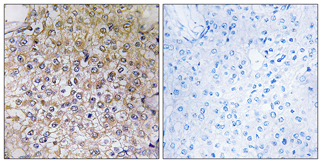
ApplicationsELISA, ImmunoHistoChemistry
ReactivityHuman, Mouse
- SizePrice

Product group Antibodies
Anti-ATP6V1B1 Antibody Picoband(r)A06073-2-CARRIER-FREE
ApplicationsWestern Blot, ELISA
ReactivityHuman
TargetATP6V1B1
- SizePrice
Product group Antibodies
Anti-ATP6V1B1 Antibody144-06876
ApplicationsWestern Blot
ReactivityHuman, Mouse, Rat
TargetATP6V1B1
- SizePrice
Product group Antibodies
ATP6V1B1 AntibodyCSB-PA002397ESR1HU
ApplicationsWestern Blot, ELISA, ImmunoHistoChemistry
ReactivityHuman, Mouse
TargetATP6V1B1
- SizePrice
Product group Antibodies

ATP6V1B1 AntibodyLS-C348977
ApplicationsImmunoFluorescence, Western Blot, ImmunoHistoChemistry
ReactivityHuman, Mouse, Rat
TargetATP6V1B1
- SizePrice
Product group Antibodies
References
V-ATPase B1 antibodyGTX32959
ApplicationsWestern Blot
ReactivityHuman, Mouse
TargetATP6V1B1
- SizePrice

Product group Antibodies
Anti-ATP6V1B1 AntibodyHPA031847
ApplicationsWestern Blot, ImmunoHistoChemistry
ReactivityHuman
TargetATP6V1B1
- SizePrice
Product group Antibodies
Anti-ATP6V1B1 AntibodyCAB6876
ApplicationsImmunoFluorescence, Western Blot, ELISA, ImmunoCytoChemistry
ReactivityHuman
TargetATP6V1B1
- SizePrice