


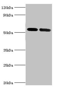
Figure 1. Western blot analysis of ATP6V1B1 using anti-ATP6V1B1 antibody (A06073-2). Electrophoresis was performed on a 5-20% SDS-PAGE gel at 70V (Stacking gel) / 90V (Resolving gel) for 2-3 hours. The sample well of each lane was loaded with 30 ug of sample under reducing conditions. Lane 1: human 293T whole cell lysates. After electrophoresis, proteins were transferred to a nitrocellulose membrane at 150 mA for 50-90 minutes. Blocked the membrane with 5% non-fat milk/TBS for 1.5 hour at RT. The membrane was incubated with rabbit anti-ATP6V1B1 antigen affinity purified polyclonal antibody (Catalog # A06073-2) at 0.5 microg/mL overnight at 4°C, then washed with TBS-0.1%Tween 3 times with 5 minutes each and probed with a goat anti-rabbit IgG-HRP secondary antibody at a dilution of 1:5000 for 1.5 hour at RT. The signal is developed using an Enhanced Chemiluminescent detection (ECL) kit (Catalog # EK1002) with Tanon 5200 system. A specific band was detected for ATP6V1B1 at approximately 69 kDa. The expected band size for ATP6V1B1 is at 57 kDa.
Figure 1. Western blot analysis of ATP6V1B1 using anti-ATP6V1B1 antibody (A06073-2). Electrophoresis was performed on a 5-20% SDS-PAGE gel at 70V (Stacking gel) / 90V (Resolving gel) for 2-3 hours. The sample well of each lane was loaded with 30 ug of sample under reducing conditions. Lane 1: human 293T whole cell lysates. After electrophoresis, proteins were transferred to a nitrocellulose membrane at 150 mA for 50-90 minutes. Blocked the membrane with 5% non-fat milk/TBS for 1.5 hour at RT. The membrane was incubated with rabbit anti-ATP6V1B1 antigen affinity purified polyclonal antibody (Catalog # A06073-2) at 0.5 microg/mL overnight at 4°C, then washed with TBS-0.1%Tween 3 times with 5 minutes each and probed with a goat anti-rabbit IgG-HRP secondary antibody at a dilution of 1:5000 for 1.5 hour at RT. The signal is developed using an Enhanced Chemiluminescent detection (ECL) kit (Catalog # EK1002) with Tanon 5200 system. A specific band was detected for ATP6V1B1 at approximately 69 kDa. The expected band size for ATP6V1B1 is at 57 kDa.
Anti-ATP6V1B1 Antibody Picoband(r)
A06073-2-CARRIER-FREE
ApplicationsWestern Blot, ELISA
Product group Antibodies
ReactivityHuman
TargetATP6V1B1
Overview
- SupplierBoster Bio
- Product NameAnti-ATP6V1B1 Antibody Picoband(r)
- Delivery Days Customer9
- ApplicationsWestern Blot, ELISA
- CertificationResearch Use Only
- ClonalityPolyclonal
- Concentration500 ug/ml
- Gene ID525
- Target nameATP6V1B1
- Target descriptionATPase H+ transporting V1 subunit B1
- Target synonymsATP6B1, DRTA2, RTA1B, VATB, VMA2, VPP3, V-type proton ATPase subunit B, kidney isoform, ATPase, H+ transporting, lysosomal 56/58kDa, V1 subunit B1, H(+)-transporting two-sector ATPase, 58kD subunit, H+-ATPase beta 1 subunit, V-ATPase B1 subunit, V-ATPase subunit B 1, endomembrane proton pump 58 kDa subunit, vacuolar proton pump 3, vacuolar proton pump subunit B 1, vacuolar proton pump, subunit 3
- HostRabbit
- Protein IDP15313
- Protein NameV-type proton ATPase subunit B, kidney isoform
- Scientific DescriptionBoster Bio Anti-ATP6V1B1 Antibody Picoband® catalog # A06073-2. Tested in WB, ELISA applications. This antibody reacts with Human. The brand Picoband indicates this is a premium antibody that guarantees superior quality, high affinity, and strong signals with minimal background in Western blot applications. Only our best-performing antibodies are designated as Picoband, ensuring unmatched performance.
- ReactivityHuman
- Storage Instruction-20°C,2°C to 8°C
- UNSPSC12352203
Related products
Product group Antibodies
Anti-ATP6V1B1 AntibodyA97821
ApplicationsELISA, ImmunoHistoChemistry
ReactivityHuman, Mouse
- SizePrice
Product group Antibodies
Anti-ATP6V1B1 Antibody144-06876
ApplicationsWestern Blot
ReactivityHuman, Mouse, Rat
TargetATP6V1B1
- SizePrice
Product group Antibodies
ATP6V1B1 AntibodyCSB-PA002397ESR1HU
ApplicationsWestern Blot, ELISA, ImmunoHistoChemistry
ReactivityHuman, Mouse
TargetATP6V1B1
- SizePrice
Product group Antibodies

ATP6V1B1 AntibodyLS-C348977
ApplicationsImmunoFluorescence, Western Blot, ImmunoHistoChemistry
ReactivityHuman, Mouse, Rat
TargetATP6V1B1
- SizePrice
Product group Antibodies
V-ATPase B1 antibody [N1C1-2]GTX110341
ApplicationsWestern Blot
ReactivityHuman
TargetATP6V1B1
- SizePrice

Product group Antibodies
Anti-ATP6V1B1 AntibodyHPA031847
ApplicationsWestern Blot, ImmunoHistoChemistry
ReactivityHuman
TargetATP6V1B1
- SizePrice
Product group Antibodies
Anti-ATP6V1B1 AntibodyCAB6876
ApplicationsImmunoFluorescence, Western Blot, ELISA, ImmunoCytoChemistry
ReactivityHuman
TargetATP6V1B1
- SizePrice