

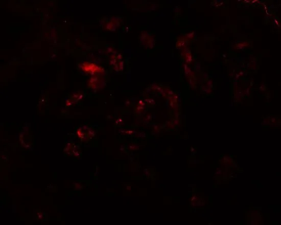
IHC-P analysis of human lung tissue using GTX17175 VAMP7 antibody. Working concentration : 20 μg/ml
 0.5 and (B) 1 μg/ml")
IHC-P analysis of human lung tissue using GTX17175 VAMP7 antibody. Working concentration : 20 μg/ml
VAMP7 antibody
GTX17175
ApplicationsWestern Blot, ELISA, ImmunoHistoChemistry, ImmunoHistoChemistry Paraffin
Product group Antibodies
ReactivityHuman, Mouse, Rat
TargetVAMP7
Overview
- SupplierGeneTex
- Product NameVAMP7 antibody
- Delivery Days Customer9
- Application Supplier NoteWB: 0.5 - 1 microg/mL. IHC-P: 20 microg/mL. *Optimal dilutions/concentrations should be determined by the researcher.Not tested in other applications.
- ApplicationsWestern Blot, ELISA, ImmunoHistoChemistry, ImmunoHistoChemistry Paraffin
- CertificationResearch Use Only
- ClonalityPolyclonal
- Concentration1 mg/ml
- ConjugateUnconjugated
- Gene ID6845
- Target nameVAMP7
- Target descriptionvesicle associated membrane protein 7
- Target synonymsSYBL1, TI-VAMP, TIVAMP, VAMP-7, vesicle-associated membrane protein 7, synaptobrevin-like 1, synaptobrevin-like protein 1, tetanus neurotoxin-insensitive VAMP, tetanus-insensitive VAMP
- HostRabbit
- IsotypeIgG
- Protein IDP51809
- Protein NameVesicle-associated membrane protein 7
- Scientific DescriptionThis gene encodes a transmembrane protein that is a member of the soluble N-ethylmaleimide-sensitive factor attachment protein receptor (SNARE) family. The encoded protein localizes to late endosomes and lysosomes and is involved in the fusion of transport vesicles to their target membranes. Alternate splicing results in multiple transcript variants.[provided by RefSeq, Jun 2010]
- ReactivityHuman, Mouse, Rat
- Storage Instruction-20°C or -80°C,2°C to 8°C
- UNSPSC41116161
References
- Unconventional secretion of FABP4 by endosomes and secretory lysosomes. Villeneuve J et al., 2018 Feb 5, J Cell BiolRead this paper
Datasheet
Related products
Product group Antibodies
VAMP7 AntibodyCSB-PA025785LA01HU
ApplicationsImmunoFluorescence, Western Blot, ELISA, ImmunoHistoChemistry
ReactivityHuman, Mouse
TargetVAMP7
- SizePrice
Product group Antibodies
Anti-VAMP7 AntibodyA49522
ApplicationsWestern Blot, ELISA
ReactivityHuman, Mouse, Rat
- SizePrice

Product group Antibodies
Anti-VAMP7 AntibodyHPA036733
ApplicationsImmunoCytoChemistry, ImmunoHistoChemistry
ReactivityHuman
TargetVAMP7
- SizePrice
Product group Antibodies

VAMP7 / SYBL1 / T1 VAMP AntibodyLS-C285636
ApplicationsWestern Blot, ELISA, ImmunoHistoChemistry
ReactivityChicken
TargetVAMP7
- SizePrice
Product group Antibodies
VAMP7 Polyclonal AntibodyCAC13995
ApplicationsImmunoFluorescence, Western Blot, ELISA, ImmunoHistoChemistry
ReactivityMouse
TargetVAMP7
- SizePrice

Product group Antibodies
Anti-SYBL1/VAMP7 Antibody Picoband(r)PB10017-CARRIER-FREE
ApplicationsWestern Blot
ReactivityBovine, Equine, Human, Monkey, Rabbit, Rat
TargetVAMP7
- SizePrice
Product group Antibodies
Anti-VAMP7 (Center) Antibody102-21624
ApplicationsWestern Blot, ImmunoHistoChemistry, ImmunoHistoChemistry Paraffin
TargetVAMP7
- SizePrice
Product group Antibodies
SYBL1 Polyclonal AntibodyBS-12852R
ApplicationsImmunoFluorescence, Western Blot, ELISA, ImmunoCytoChemistry, ImmunoHistoChemistry, ImmunoHistoChemistry Frozen, ImmunoHistoChemistry Paraffin
ReactivityBovine, Chicken, Equine, Human, Mouse, Porcine, Rabbit, Rat, Sheep, Zebra Fish
TargetVAMP7
- SizePrice